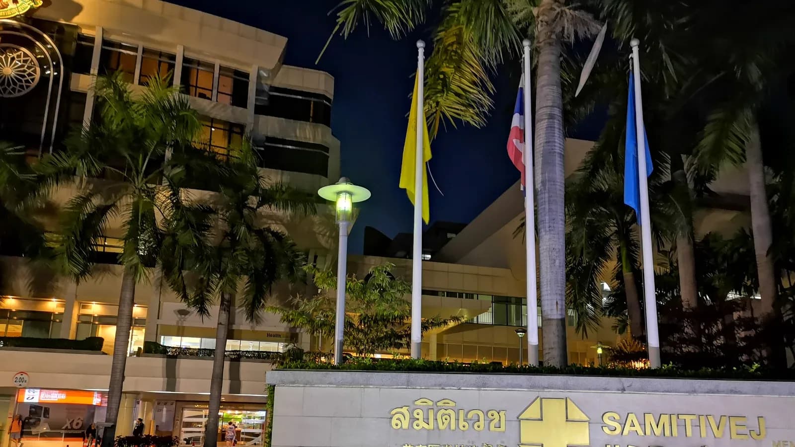
Samitivej Sukhumvit Hospital
bangkok
- Specialties
- 26
- Departments
- 29
Not sure which hospital fits your case?
Upload your medical records and let AI match you to the right hospital.
Upload records and get matchedAbout
Samitivej Sukhumvit Hospital, on Sukhumvit Road in Bangkok, is a leading Joint Commission International accredited hospital and part of the Bangkok Dusit Medical Services (BDMS) group, founded in 1979. With more than 400 beds, it is especially well known for paediatrics and children's care, women's health and a large international-patient programme, and provides multi-super-specialty care across cardiac sciences, cancer, neuroscience, orthopaedics and spine, organ transplantation, fertility and robotic surgery.
Specialties
Departments
- Cardiac Sciences
- Medical Oncology
- Radiation Oncology
- Breast Health
- Hematology and Bone Marrow Transplant
- Neurosciences
- Neurosurgery
- Spine Surgery
- Gastroenterology
- Gastrointestinal Surgery
- Orthopaedics and Joint Replacement
- Urology
- Nephrology
- Organ Transplantation
- Liver Transplant
- Pulmonology
- Obstetrics and Gynaecology
- Reproductive Medicine and Fertility
- Paediatrics
- General Surgery
- Robotic Surgery
- Plastic and Reconstructive Surgery
- Dermatology
- Endocrinology and Diabetes Care
- Ophthalmology
- ENT
- Rheumatology
- Physiotherapy and Rehabilitation
- Preventive Health and Check-up
Procedures
Cardiology and Cardiovascular Surgery
Ear, Nose and Throat
General Surgery
Gynecology and Obstetrics
IVF and Reproductive Health
Neurology and Neurosurgery
Oncology
Organ Transplantation
Orthopedics and Traumatology
Physical Medicine and Rehabilitation
Plastic and Aesthetic Surgery
International patient services
- International patient office
- Interpreter and translation services
- Visa and travel assistance
- Airport transfer
- Accommodation assistance
Technologies and equipment
CyberKnife M6
CyberKnife M6 is a robotic system for stereotactic radiosurgery and stereotactic body radiotherapy (SBRT). Despite the name, there is no knife and no cutting. A small linear accelerator sits on a computer-guided robotic arm and delivers many thin beams of focused radiation from hundreds of angles. The beams converge on the tumour with sub-millimetre accuracy, so a high dose reaches the target while nearby healthy tissue is spared. Imaging during treatment tracks the tumour continuously, and a motion-synchronisation feature follows targets that move with breathing, such as those in the lung or liver. Treatment is non-invasive and painless, needs no rigid head frame, and is usually given as an outpatient over one to five sessions. The decision is always made individually by the radiation oncology team.
PET-CT
PET-CT is an advanced hybrid imaging method that combines positron emission tomography with computed tomography in a single scan, mapping both the metabolic activity and the anatomical structure of the body at once. A small dose of a radioactive tracer, often a glucose analogue, is injected and gathers in cells that are working harder than normal, which is typical of many tumours. Because it can show where a disease is active before it changes the shape of an organ, PET-CT is one of the most valuable tools for detecting cancer, working out how far it has spread, and checking whether treatment is working.
Da Vinci Robotic Surgery
The da Vinci robotic surgical system lets a surgeon perform complex operations through a few small keyhole incisions instead of one large cut. Sitting at a nearby console, the surgeon controls tiny wristed instruments and a magnified high-definition three-dimensional camera, while the robotic arms translate every hand movement into precise, steady motion inside the body. The system never acts on its own: the surgeon is in full control at all times. For patients, this minimally invasive approach often means less pain, smaller scars, less blood loss and a quicker return to normal life.
3 Tesla MRI
3 Tesla MRI is a high-field magnetic resonance imaging scanner that produces exceptionally detailed pictures of the inside of the body. The "3 Tesla" refers to the strength of its magnet, which is about twice that of a standard MRI scanner, and this extra power allows sharper, higher-resolution images, often in less time. Like all MRI, it uses a strong magnetic field and radio waves rather than X-rays, so there is no ionising radiation involved. It is especially valuable for examining the brain, the nervous system, joints and soft tissues, helping doctors detect and characterise problems that may be hard to see on other scans.
Digital Mammography
Digital mammography is a low-dose X-ray method used to screen for and detect breast cancer at an early stage. It captures very high-resolution digital images of the breast that a radiologist can examine and enhance on screen, revealing small nodules, masses and tiny specks of calcium that may not be felt or seen on other tests. Because it can find changes long before they cause symptoms, it is the cornerstone of breast cancer screening and one of the most effective tools for catching the disease when it is most treatable.
Robotic Arm-Assisted Orthopedic Surgery
Robotic arm-assisted orthopedic surgery is a technology used mainly in knee and hip replacement to plan and carry out the operation with very high accuracy. A detailed three-dimensional plan is built from the patient's own CT scan, and during surgery a robotic arm guides the surgeon's instruments so that bone is prepared and the implant is positioned to that exact plan. The surgeon always holds and directs the instrument; the robotic arm adds steadiness and built-in limits that protect the surrounding tissue. The aim is a joint that fits and balances well, which can mean less pain and a smoother recovery.
Location
133 Sukhumvit 49, Klongtan Nua, Wattana, Bangkok 10110, Thailand
View on Google MapsAccreditations
- JCI
Not sure which hospital fits your case?
Upload your medical records and let AI match you to the right hospital.
Upload records and get matched