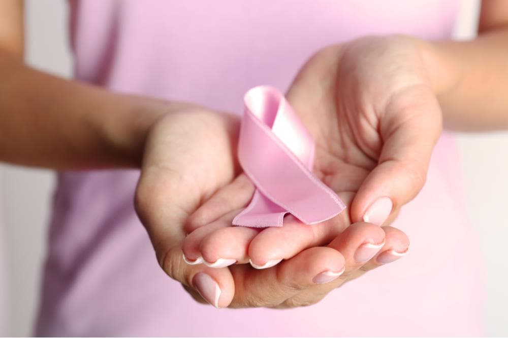
Breast Health

Breast Health

Gebze

Breast Health care in Gebze is available at 1 hospital in the Voumed network.

Breast health care looks after the whole range of breast conditions, from harmless lumps and cysts to breast cancer, with one central aim: to find any serious disease early, when it is most treatable. Breast cancer is among the most common cancers in women, yet when it is caught while still small and confined to the breast, treatment is usually very successful. People travel abroad for breast care to reach experienced teams, modern imaging and the multidisciplinary approach that brings surgeons, oncologists, radiologists and pathologists together around a single plan. A dedicated breast service offers not only diagnosis and treatment but also reassurance, since most breast changes turn out to be benign.

On this page

At a glance

Sub-specialties
breast surgery, breast imaging and radiology, medical oncology, radiation oncology, pathology, oncoplastic and reconstructive surgery
Common procedures
clinical examination, mammography, breast ultrasound, breast MRI, needle biopsy, breast-conserving and reconstructive surgery
Common reasons to travel
experienced multidisciplinary teams, advanced imaging, integrated cancer care and the option of a second opinion
Typical hospital stay
outpatient for screening, diagnosis and biopsy, day case or 1 to 3 nights for breast surgery
Anaesthesia
local anaesthesia for most biopsies, general anaesthesia for breast surgery
Typical first step
a consultation with a clinical breast examination and imaging chosen for the individual

Overview

Breast health combines screening, diagnosis and treatment in one connected pathway. Screening looks for disease before it causes symptoms, diagnosis works out what a lump or an imaging finding actually is, and treatment is tailored to the result. The single most important idea is early detection: breast cancer found while it is still a small mass inside the breast, before it spreads through the blood and lymph, can usually be treated with full success. Because early disease often causes no symptoms at all, regular screening and the prompt assessment of any change are what make the difference. A modern breast centre is built around a team, so that imaging, biopsy and, if needed, treatment can move forward quickly and without fragmentation.

Conditions and sub-specialties

Most breast conditions are benign. These include simple cysts, fibroadenomas, which are smooth benign lumps common in younger women, fibrocystic change, breast pain and benign nipple discharge, all of which can be diagnosed and reassured or treated as needed. Breast cancer is the central malignant condition, and it ranges from very early non-invasive disease confined to the milk ducts to invasive cancer that needs fuller treatment. Some people carry an inherited risk, for example through a family history or a known gene change, and benefit from earlier and more intensive screening and from genetic risk assessment. The breast service draws on several specialties: breast surgery, breast imaging, medical and radiation oncology, pathology and reconstructive surgery, each contributing to the diagnosis and the plan.

Common treatments and procedures

Assessment begins with a clinical examination and imaging, and if a finding needs to be clarified, a needle biopsy under local anaesthesia gives a definitive answer, usually as an outpatient. When cancer is confirmed, treatment is planned by the team and may combine several approaches. Surgery may remove only the lump and a margin around it, known as breast-conserving surgery, or the whole breast when that is more appropriate, often with the option of reconstruction at the same time or later. Medical treatments such as chemotherapy, hormone therapy or targeted therapy, and radiation therapy, are added according to the type and stage of the disease. For benign conditions, treatment may be as simple as reassurance and follow-up, the drainage of a cyst, or the removal of a benign lump if it is troublesome.

Diagnostics and technology

Breast imaging is the cornerstone of early detection. Mammography, a low dose X-ray of the breast, is the main screening tool and can show very small changes before they can be felt; modern digital and tomosynthesis, or three dimensional, mammography improves the view of dense breast tissue. Breast ultrasound helps tell solid lumps from fluid-filled cysts and guides biopsies, and breast MRI is used in selected situations such as high inherited risk or dense tissue. When imaging finds something that needs clarifying, an image-guided needle biopsy takes a small tissue sample for the pathologist, who provides the definitive diagnosis and the detailed information that guides treatment. Used together, these tools detect disease early, separate benign conditions from cancer and match each patient with the right plan.

What to expect as an international patient

For people who travel, care usually begins with a remote consultation and a review of any mammograms, scans or reports already done, which can save time on arrival. A focused visit can then combine examination, imaging and, where needed, a biopsy within a short stay, often a few days, with pathology results following soon after. If treatment is required, the length of stay depends on the plan: surgery may mean a day case or one to three nights, while courses of chemotherapy or radiation therapy may be arranged to suit travel, sometimes with parts continued nearer home. Your team will advise when it is safe to fly after surgery. Interpreter and international patient support helps with appointments, consent and understanding the results, and a second opinion on an existing diagnosis is a common reason to travel.

Frequently asked questions

These answers are general guidance and may vary by provider. Confirm the details with the hospital you choose.

What does a breast health centre do?

A breast health centre covers screening, diagnosis and treatment for the whole range of breast conditions, from benign lumps and cysts to breast cancer. It combines clinical examination, imaging and biopsy with a multidisciplinary team that plans any treatment together.

Why is early detection of breast cancer so important?

Breast cancer found while it is still a small mass inside the breast, before it spreads through the blood and lymph, can usually be treated with full success. Because early disease often causes no symptoms, regular screening and the prompt assessment of any change give the best chance of cure.

How is a breast problem diagnosed?

Diagnosis combines a clinical examination with imaging such as mammography, ultrasound and, when needed, MRI. If a finding needs clarifying, an image-guided needle biopsy under local anaesthesia gives a definitive answer and tells benign conditions apart from cancer.

I have found a lump. Does it mean cancer?

Not usually. Most breast lumps are benign, such as cysts or fibroadenomas, but any new lump or change in the breast should be assessed promptly so it can be identified with certainty and treated if needed.

How is breast cancer treatment decided?

A patient with breast cancer is assessed by a multidisciplinary team in which breast surgery, medical and radiation oncology, radiology and pathology review the case together and agree an individualised plan, with reconstruction considered as part of it.

Can I come for a second opinion on a breast cancer diagnosis?

Yes. Many people travel specifically for a second opinion. The team can review your existing imaging, biopsy and pathology, repeat tests if needed and confirm or refine the diagnosis and treatment plan.

How many days should I plan to stay abroad?

For screening, diagnosis and biopsy, a stay of a few days is often enough, with pathology results following soon after. If surgery or other treatment is needed, the stay is longer and depends on the plan, which your team will set out clearly in advance.

Will there be interpreter or language support?

Yes. International patient services commonly include interpreters and coordinators who help with appointments, consent, understanding the results and day to day questions, so language is not a barrier to safe care.

Not sure which hospital fits your case?

Upload your medical records and let AI match you to the right hospital.

Upload records and get matched

Available at these hospitals

Technologies and equipment

4D Breast Ultrasound

4D breast ultrasound, also known as automated breast volumetric scanning, is an imaging method that supports the diagnosis of breast cancer. Unlike a hand-held ultrasound, it uses a dedicated probe that moves automatically across the breast to capture the whole organ as a complete three-dimensional volume. It is mainly used alongside mammography, especially for women with dense breast tissue, where a denser background can hide lesions on a standard mammogram. The examination is comfortable, non-invasive and free of ionising radiation, making it a valuable additional layer in breast screening and assessment.

View technology

Intraoperative Radiotherapy (IORT)

Intraoperative radiotherapy, or IORT, is a way of giving a single, focused dose of radiation directly to the area at highest risk during the operation itself, right after the tumour has been removed and while the patient is still asleep under anaesthesia. With the surgical wound open, the team can place the radiation applicator exactly on the tissue that needs it and gently move skin and nearby organs out of the path of the beam. The result is treatment that is aimed precisely where cancer cells are most likely to remain, while healthy tissue is shielded. For suitable patients, especially in early breast cancer, one intraoperative dose can replace or greatly shorten several weeks of radiotherapy after surgery.

View technology

Digital Mammography

Digital mammography is a low-dose X-ray method used to screen for and detect breast cancer at an early stage. It captures very high-resolution digital images of the breast that a radiologist can examine and enhance on screen, revealing small nodules, masses and tiny specks of calcium that may not be felt or seen on other tests. Because it can find changes long before they cause symptoms, it is the cornerstone of breast cancer screening and one of the most effective tools for catching the disease when it is most treatable.

View technology

Tomosynthesis Mammography (3D Mammography)

Tomosynthesis mammography, often called 3D mammography, is an advanced form of digital mammography that builds a three-dimensional picture of the breast from a series of thin layers. Instead of a single flat image in which overlapping tissue can hide or mimic a problem, it lets the radiologist scroll through the breast slice by slice on a high-resolution screen. This makes small lesions and tumours easier to see and helps distinguish real findings from harmless overlapping tissue, which is especially valuable for screening and for women with dense breasts.

View technology

Not sure which hospital fits your case?

Upload your medical records and let AI match you to the right hospital.

Upload records and get matched