How a Colonoscopy Is Performed: A Complete Beginner's Guide
A colonoscopy is an outpatient endoscopic test examining the large intestine to detect and remove precancerous polyps under sedation in 15 to 45 minutes.

How a Colonoscopy Is Performed: A Complete Beginner's Guide
Quick Answer: A colonoscopy is an outpatient endoscopic procedure that examines the mucosal lining of the large intestine to detect inflammation, ulcers, and precancerous polyps. Performed under gentle intravenous sedation in 15 to 45 minutes, it allows immediate removal of polyps, helping prevent colorectal cancer before symptoms develop.
Key Takeaways:
- Routine screening begins at age 45 for average-risk adults to identify and remove precancerous growths early.
- Gastroscopy and colonoscopy are frequently performed in a single session under one sedation cycle to evaluate the entire digestive tract.
- Bowel preparation requires a short transition to a low residue diet followed by clear liquids and a prescribed laxative solution.
- The visual examination and polyp removal cause no pain due to modern sedation and the absence of sensory pain receptors in the colonic mucosa.
- Most patients leave the clinic within two to three hours of arrival and resume normal eating the same day.
A colonoscopy is a direct visual examination of the large bowel (colon and rectum) using a thin, flexible endoscope equipped with digital optics and working channels. As both an exploratory diagnostic test and a preventative therapeutic procedure, it serves as the clinical standard for detecting mucosal abnormalities, stopping gastrointestinal bleeding, and removing abnormal tissue before it can transform into invasive cancer.
What is a colonoscopy and why is it performed?
A colonoscopy is an endoscopic examination where a specialist uses a flexible, illuminated tube equipped with a camera (colonoscope) to inspect the mucosal lining of the rectum and entire large intestine. Physicians use it to investigate unexplained gastrointestinal symptoms, detect inflammation, and identify precancerous growths before they progress.
Beyond screening asymptomatic adults, doctors recommend the procedure to evaluate symptoms that could indicate lower gastrointestinal pathology. These clinical indications include:
- Persistent abdominal pain, cramping, or unexplained bloating
- Chronic diarrhea, constipation, or sudden changes in bowel habits
- Unexplained iron deficiency anemia or chronic fatigue
- Visible rectal bleeding or dark, tarry stools
- Routine surveillance for patients with a personal history of inflammatory bowel disease (Crohn's disease or ulcerative colitis)
Unlike radiological imaging (such as CT colonography) or non-invasive stool tests (such as fecal immunochemical tests), a colonoscopy offers an immediate therapeutic advantage. When abnormal tissue or bleeding vessels appear during the scan, the specialist passes miniature instruments directly through the scope's working channel to take tissue samples or excise growths in real time.
Why doctors often combine colonoscopy with gastroscopy in a single session
Dual endoscopy combines an upper gastrointestinal examination (gastroscopy) and a lower bowel examination (colonoscopy) during a single sedation session. This simultaneous approach allows clinicians to evaluate both upper tract symptoms (such as reflux, ulcers, and stomach inflammation) and lower bowel pathology without subjecting the patient to two separate hospital visits or multiple rounds of sedation.
An upper endoscopy, also called an esophagogastroduodenoscopy (EGD) or gastroscopy, uses a narrower endoscope inserted through the mouth to visualize the esophagus, stomach, and duodenum. Combining both examinations is common when patients present with complex symptoms that could originate in either region, such as systemic iron deficiency, unintended weight loss, or generalized abdominal discomfort.
The dual approach offers distinct clinical advantages:
- Single Sedation Event: Patients receive intravenous sedation only once, minimizing anesthetic drug exposure and post-procedure grogginess.
- Unified Fasting Window: Pre-procedure dietary restrictions and fasting protocols cover both examinations simultaneously.
- Comprehensive Diagnostic Yield: Clinicians obtain a full diagnostic assessment from the upper digestive tract down to the ileocecal junction in under an hour.
- Logistical Efficiency: Patients arrange transportation, time off work, and clinic recovery for a single day rather than two separate appointments.
Colonoscopy age guidelines: When should you schedule your first screening?
Major gastroenterology guidelines recommend that average-risk individuals begin colonoscopy for colon cancer screening at age 45. Individuals with a family history of colorectal neoplasms, genetic syndromes like Lynch syndrome, or chronic inflammatory bowel disease typically begin screening earlier, often at age 40 or 10 years younger than the earliest diagnosed first-degree relative.
The standard colonoscopy age baseline was lowered from 50 to 45 by major medical societies due to rising rates of early-onset colorectal neoplasia in younger adults. Routine screening remains recommended through age 75, with decisions between ages 76 and 85 individualized based on overall health status, prior screening findings, and life expectancy.
| Risk Category | Criteria | Recommended Starting Age | Standard Interval |
|---|---|---|---|
| Average Risk | No personal/family history of polyps or colorectal cancer; no IBD | Age 45 | Every 10 years |
| Increased Familial Risk | First-degree relative diagnosed with colorectal cancer before age 60 | Age 40, or 10 years earlier than relative's diagnosis | Every 5 years |
| High Genetic Risk | Confirmed Lynch syndrome or familial adenomatous polyposis | Age 20-25 (or earlier based on gene variant) | Every 1-2 years |
| Inflammatory Bowel Disease | Longstanding ulcerative colitis or Crohn's colitis | 8 years after symptom onset | Every 1-3 years |
How to prepare: Low residue diet, clear liquids, and the colonoscopy cleanse
Optimal bowel cleansing requires transitioning to a low residue diet for colonoscopy 3 to 5 days before the appointment, shifting to a clear liquid diet 24 hours prior, and completing the prescribed osmotic or polyethylene glycol laxative regimen. Complete adherence ensures the mucosal wall remains free of fecal debris so the endoscopist can detect even flat or miniature lesions.
The diagnostic accuracy of the examination directly correlates with mucosal visibility. Residual stool can obscure small adenomas, leading to missed lesions or requiring an early repeat examination. The bowel preparation protocol occurs in progressive phases:
| Timeline | Permitted Nutrition | Items to Avoid | Preparation Actions |
|---|---|---|---|
| Days 3 to 2 Before | White bread, white rice, poultry, eggs, peeled potatoes | Whole grains, nuts, seeds, raw vegetables, beans, corn | Discontinue iron supplements and high-fiber bulk additives |
| Day 1 Before | Clear broth, water, apple juice, plain gelatin, black coffee | All solid food, dairy, alcohol, red or purple dyed liquids | Start clear liquid diet for colonoscopy; drink first prep dose |
| Examination Day | Clear water only up to 2-4 hours before arrival | All food, drinks, mints, and chewing gum | Drink second prep dose (split-dose); maintain strict fasting |
During the low residue phase, eliminating fiber reduces the volume of undigested matter traveling through the colon. On the final preparation day, the clear liquid diet prevents new solid waste from forming.
The colonoscopy cleanse solution is typically taken as a split dose: the patient drinks the first half the evening before and the second half 4 to 6 hours before the examination. Split dosing consistently yields superior bowel cleanliness compared to single-day dosing.
Step-by-step: How the procedure is carried out on examination day
On procedure day, medical staff administer intravenous conscious or deep sedation, position the patient on their left side, and gently advance the colonoscope through the rectum to the cecum while inflating the lumen with carbon dioxide or air. If the endoscopist identifies suspicious tissue or colonoscopy polyps, they excise them immediately with specialized snare tools (polypectomy) for pathology analysis without causing pain.
The procedure follows a standardized clinical workflow:
- Pre-Procedure Assessment: Nursing staff verify vital signs, confirm fasting compliance, and insert a small intravenous (IV) catheter into your arm or hand.
- Sedation Administration: An anesthesiologist or sedation-certified physician administers fast-acting intravenous medications (typically propofol or midazolam with fentanyl). You drift into a restful sleep within seconds while breathing comfortably on your own.
- Endoscope Insertion: The specialist lubricates the tip of the colonoscope and gently inserts it through the rectum, navigating through the colon to reach the cecum (the junction with the small intestine). Carbon dioxide is gently infused to expand the intestinal walls for clear visualization.
- Careful Mucosal Inspection: The critical inspection occurs during the slow withdrawal of the instrument, which takes at least 6 to 10 minutes. The gastroenterologist inspects the entire mucosal surface under high-definition white light and digital chromoendoscopy.
- Polyp Removal and Biopsy: If the physician discovers abnormal mucosal growths or colonoscopy polyps, they advance miniature surgical tools through the scope's internal channel. Cold snares or biopsy forceps remove smaller polyps mechanically, while electrosurgical snares use low-voltage current to resect larger vascular polyps. Because the inner lining of the colon contains no pain receptors, these interventions cause no discomfort.
How long does colonoscopy take and what happens during recovery?
The diagnostic examination itself typically takes 15 to 45 minutes, with total facility time averaging 2 to 3 hours to account for pre-procedure preparation and post-sedation recovery. Patients wake up within 30 minutes in a recovery bay, may experience mild temporary bloating from residual gas, and generally resume normal eating later that day once discharge criteria are met.
If no polyps are found, the procedure usually finishes in approximately 20 minutes. If multiple large polyps require removal or if a combined gastroscopy is performed, the total procedure time may extend toward 45 to 60 minutes.
Post-examination recovery involves several standard steps:
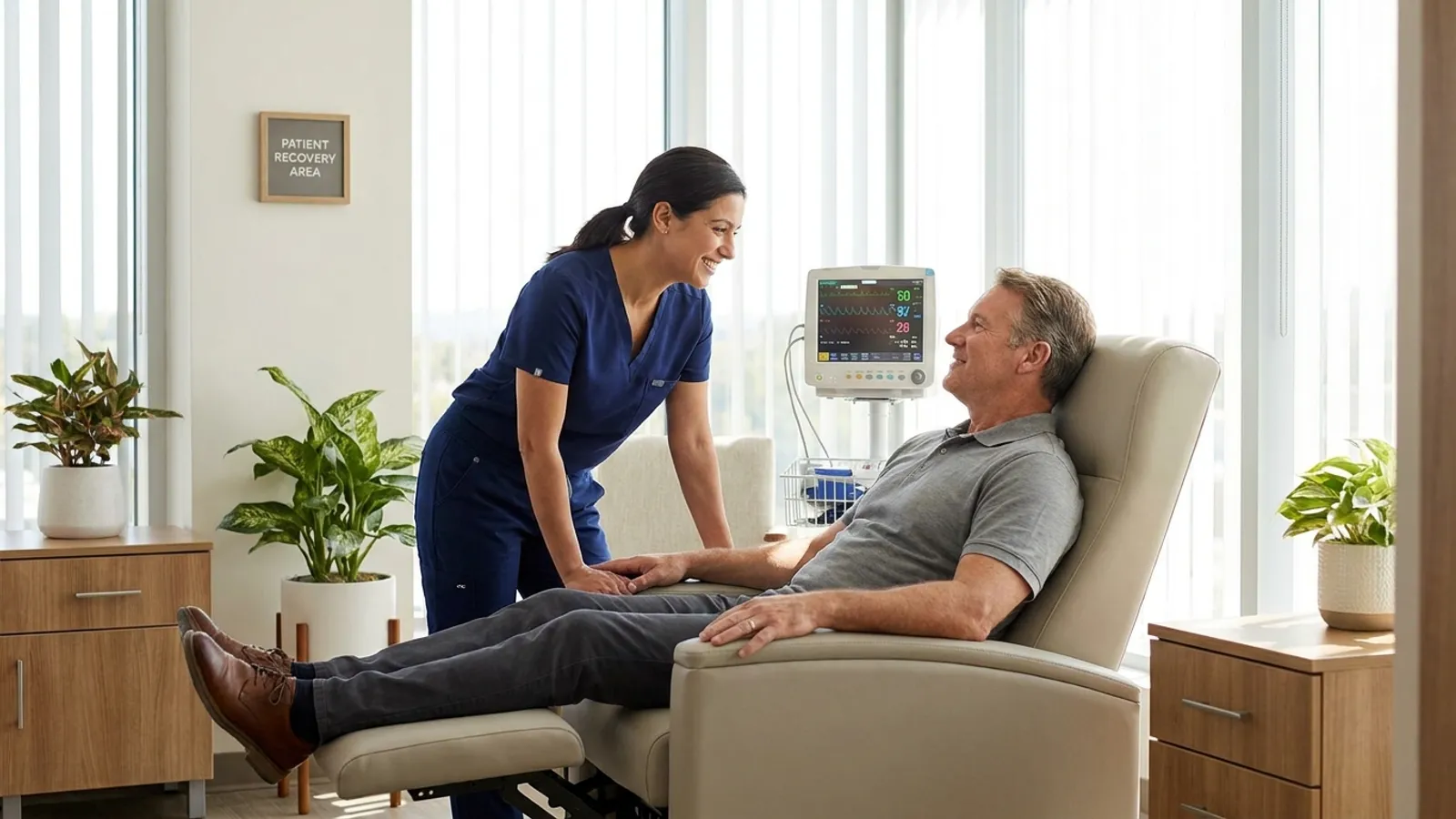
- Immediate Recovery (30-45 minutes): Sedation wears off rapidly. Clinical staff monitor vital signs until you are alert and oriented.
- Managing Intestinal Gas: Patients may feel mild cramping or fullness from residual gas. Modern clinics use medical carbon dioxide instead of room air for insufflation because carbon dioxide absorbs into the bloodstream up to 150 times faster, significantly minimizing post-procedure bloating.
- Dietary Resumption: Once awake and tolerating fluids, patients can eat a light meal (such as toast, soup, or crackers), progressing to a normal diet as comfort allows.
- Discharge Guidelines: Because sedative medications subtly impair cognitive processing and motor reflexes, an adult companion must escort you home. Patients must not drive, operate machinery, sign legal documents, or consume alcohol for 24 hours post-procedure.
Understanding your colonoscopy results: Polyps, biopsies, and follow-up intervals
A normal examination with clear visualization typically warrants a repeat screening in 10 years for average-risk individuals. When polyps are removed, histological analysis determines whether they are benign hyperplastic growths or precancerous adenomas, which guides whether the patient returns for surveillance in 3, 5, or 7 years based on polyp size and count.
Before discharge, the gastroenterologist provides an initial verbal summary of what was observed. Any removed tissue is sent to a pathology laboratory for microscopic analysis, with formal histology reports typically available within 5 to 10 business days.
Pathology findings generally fall into specific histological classifications:
- Hyperplastic Polyps: Small, non-cancerous growths primarily located in the rectum and lower colon that do not increase cancer risk.
- Tubular Adenomas: The most common type of precancerous polyp. Removing them completely prevents their potential transformation into malignancy.
- Sessile Serrated Lesions (SSLs): Flat, subtle polyps typically found in the right colon that require careful resection and structured follow-up.
- Tubulovillous or Villous Adenomas: Growths with finger-like microscopic projections that carry a higher risk of cellular dysplasia, warranting shorter surveillance intervals.
| Histology / Exam Finding | Clinical Classification | Recommended Surveillance Interval |
|---|---|---|
| Normal Examination | Clean mucosal wall, no polyps | 10 years |
| 1-2 Small Tubular Adenomas (<10 mm) | Low-risk adenoma | 7-10 years |
| 3-4 Small Tubular Adenomas | Intermediate-risk adenoma | 3-5 years |
| 5-10 Adenomas, or Any Polyp ≥10 mm | High-risk adenoma | 3 years |
| More than 10 Polyps on Single Exam | Multiple adenomas | 1 year (with genetic evaluation) |
| Incomplete Bowel Preparation | Suboptimal visualization | Repeat within 1 year |
Risks and potential complications to consider
A colonoscopy is a safe and routine outpatient procedure, but like any medical intervention involving instruments and sedation, it carries low-frequency risks such as bleeding, colonic perforation, or adverse drug reactions. Overall serious complication rates remain well below 0.3% in standard screening populations.
Complications are uncommon when procedures are conducted by experienced gastroenterologists in accredited medical facilities. Potential clinical risks include:
- Post-Polypectomy Bleeding: Occurs in approximately 0.5% to 1.5% of polyp removals. Bleeding can happen immediately or appear up to two weeks later as the healing site sheds its protective scab. Most minor bleeding resolves spontaneously or is managed with endoscopic clips.
- Intestinal Perforation: A small tear or puncture in the bowel wall, occurring in roughly 1 in 1,000 to 1 in 3,000 routine examinations. While rare, risk is slightly higher during the complex excision of large, flat polyps. Small perforations are often closed during the procedure using endoscopic clips, though some may require surgical repair.
- Adverse Sedation Reactions: Temporary decreases in blood pressure, shallow breathing, or mild allergic reactions occur in fewer than 0.5% of patients and respond rapidly to supplementary oxygen or medication adjustments.
- Infection: Extremely rare due to strict hospital reprocessing standards and single-use endoscopic instruments.
Patients should contact their medical team or seek emergency care immediately if they experience severe, worsening abdominal pain that does not improve after passing gas, continuous heavy rectal bleeding (passing clots or more than a few tablespoons of blood), chills, or a fever above 100.4°F (38°C).
Frequently Asked Questions
How long does colonoscopy take from arrival to departure?
The total facility time is typically 2 to 3 hours from registration to discharge. While the endoscopic examination itself requires only 15 to 45 minutes, patients spend 45 to 60 minutes on pre-procedure nursing intake and IV placement, followed by 45 to 60 minutes in the recovery bay while sedation fully clears.
Will I feel pain during a colonoscopy or polyp removal?
Patients feel no pain during the procedure because intravenous sedation maintains deep relaxation or light sleep throughout the examination. In addition, the internal lining of the large intestine does not possess somatic pain receptors, meaning tissue biopsies and polyp excisions cause no physical sensation even if you are lightly awake.
Can I take my regular medications during the bowel cleanse?
Most critical daily medications-such as those for high blood pressure, thyroid conditions, or seizures-can be taken with small sips of water on the morning of your examination. However, blood thinners, anticoagulants, diabetes medications, and oral iron supplements require individualized dosing adjustments or temporary pauses under your physician's guidance several days prior.
What happens if my bowel preparation is incomplete on the day of the exam?
If your bowel movements are not clear, yellowish liquid by the morning of the procedure, remaining stool can obstruct the camera's view of the colon wall. In this scenario, the clinical team may administer an additional in-clinic laxative dose or reschedule the test to prevent missed polyps and ensure accurate diagnostic results.
Not sure which hospital is right for your case?
Upload your medical reports and our AI will match them against the verified capabilities of every hospital in our network.
- Upload your reports (no account needed)
- Our AI matches your case with hospitals that can actually treat it
- You choose who receives your case. We present it in the hospital’s own language, and the hospital replies directly to you
Free and anonymous. Your documents stay private until you decide to send them.