Ferritin Face: Can You Spot Low Iron from Facial Appearance? Symptoms, Levels, and Clinical Tests
Facial changes cannot diagnose low ferritin. Discover true symptoms of iron deficiency, normal haemoglobin links, and UK serum ferritin guidelines.

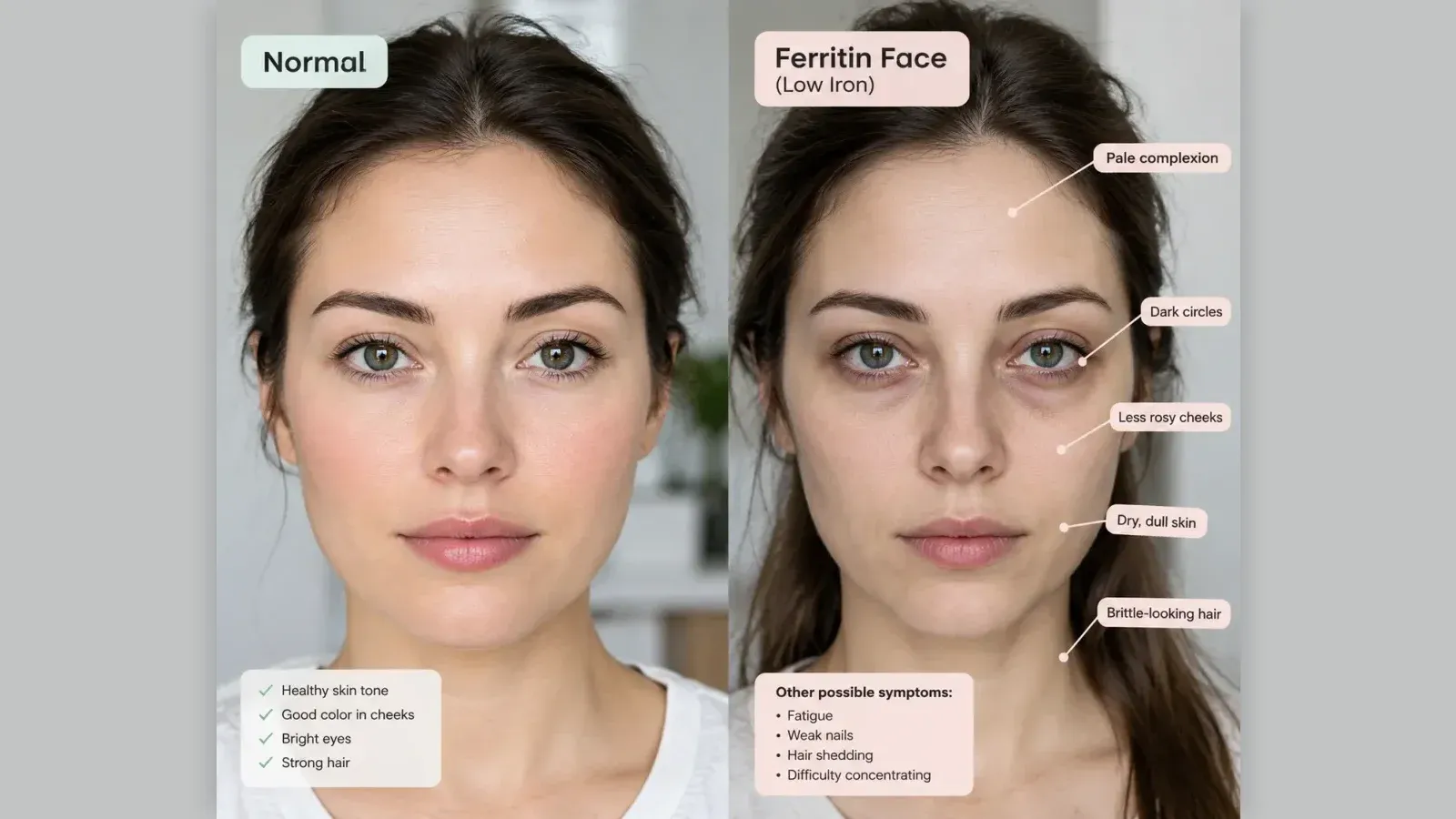
Quick Answer: Facial changes such as dull skin or under-eye dark circles cannot reliably diagnose low ferritin. A validated ferritin blood test is required to evaluate true cellular iron stores accurately.
Key Takeaways:
- The colloquial concept of "ferritin face" mistakes non-specific cosmetic changes for internal cellular iron depletion.
- Iron deficiency without anaemia occurs when stored ferritin drops while circulating haemoglobin remains within normal limits.
- Confirmed clinical signs of low ferritin include fatigue, exertional breathlessness, brain fog, and telogen effluvium hair shedding.
- UK guidelines establish that serum ferritin below 15 µg/L confirms absolute deficiency, with levels below 30 µg/L prompting clinical investigation.
- Self-treating with high-dose iron carries toxicity risks, meaning investigation of root causes must always precede therapeutic repletion.
The term "ferritin face" gained traction on social video platforms as shorthand for visible changes attributed to depleted iron reserves, including sallow complexions, persistent dark circles, and lacklustre skin. Clinically, ferritin is a globular intracellular protein complex that stores iron in a non-toxic, bioavailable form across the liver, spleen, and bone marrow. While systemic iron is fundamental to cellular oxygenation, mitochondrial energy synthesis, and collagen metabolism, external appearance alone does not provide a valid clinical marker of haematological status.
What Is 'Ferritin Face' and Can You Spot Low Iron from Facial Changes?
You cannot reliably diagnose low ferritin or iron deficiency from your face alone. While depleted iron stores can lead to subtle pallor, under-eye darkening, and diffuse hair shedding as systemic oxygen transport decreases, these cosmetic features are non-specific. They overlap heavily with genetics, fatigue, thyroid disorders, and routine skin aging. A definitive assessment requires a validated serum ferritin blood panel.
In clinical dermatology and haematology, the visible hallmarks linked online to a ferritin face lack diagnostic specificity. Sallow skin and pallor occur when tissues receive lower levels of oxygenated blood, which can prompt the microvascular capillary beds of the face to constrict. However, this physiological response is identical across several conditions, including vitamin B12 deficiency, hypothyroidism, dehydration, poor sleep, and chronic stress.
Periorbital dark circles, frequently cited on social media as proof of iron depletion, are largely dictated by:
- Craniofacial bone architecture and suborbital fat distribution.
- Microvascular pooling beneath thin lower-eyelid skin.
- Genetic post-inflammatory hyperpigmentation.
- Natural loss of periorbital collagen over time.
While severe iron deficiency anaemia can cause visible mucosal and subungual pallor on the inner lower eyelids, gums, and nail beds, early to moderate iron store depletion rarely produces distinctive facial changes. Attempting to deduce internal iron stores from cheek colour or eyelid pigmentation bypasses essential medical diagnostics. It also risks mistaking serious metabolic or endocrine imbalances for an aesthetic shortfall.
Ferritin vs Haemoglobin: Iron Deficiency Without Anaemia Explained
Ferritin measures your body's stored reserves of iron inside cells, whereas haemoglobin reflects circulating iron actively transporting oxygen in red blood cells. A patient can have low ferritin with normal haemoglobin, a state known clinically as iron deficiency without anaemia (IDWA). In this state, cellular energy production suffers long before overt anaemia registers on a routine full blood count.
Understanding the difference between functional and stored iron clarifies why standard initial tests may miss significant depletion. Haemoglobin is an iron-rich protein packed within red blood cells, functioning as the primary carrier of respiratory gases from lungs to peripheral tissues. If dietary intake or internal reserves drop, the body prioritises haemoglobin synthesis above other iron-dependent pathways to sustain immediate oxygen transport to vital organs.
Ferritin acts as the physiological buffer. When total body iron declines, cellular ferritin stores are gradually emptied to supply the bone marrow for continued red blood cell production (erythropoiesis). A routine full blood count (FBC) records haemoglobin concentration, mean corpuscular volume (MCV), and haematocrit. In early depletion, an FBC often returns entirely normal values because red blood cell production has not yet fallen.
This creates a widespread clinical presentation: low ferritin normal haemoglobin. Even though oxygen carriage remains technically intact, the depletion of non-haemoglobin iron compromises crucial intracellular processes, including:
- Cytochrome oxidase activity within the mitochondrial respiratory chain, causing cellular energy deficits.
- Myoglobin synthesis within skeletal muscle fibres, impairing physical stamina.
- Neurotransmitter synthesis, particularly dopamine and serotonin pathways dependent on iron as an essential enzymatic cofactor.
- Ribonucleotide reductase function, slowing cellular proliferation in rapidly dividing tissues such as skin and hair follicles.
Consequently, patients frequently experience debilitating physical symptoms despite their routine screening panels showing non-anaemic results.
Verified Signs and Low Ferritin Levels and Symptoms
True iron store depletion manifests primarily through systemic physiological dysfunction rather than isolated aesthetic cues. The most common verified symptoms include persistent physical fatigue, exertional breathlessness, brain fog, restless legs syndrome, cold intolerance, brittle spoon-shaped nails (koilonychia), and telogen effluvium, an accelerated shedding of scalp hair.
When iron stores dwindle, the body rations resources away from peripheral, high-turnover structures to protect critical cardiovascular and cerebral functions. The clinical presentation of low ferritin levels and symptoms reflects this cellular deprivation across multiple body systems:
- Profound, Unrefreshing Fatigue: A continuous loss of physical and cognitive energy that does not resolve after standard sleep, caused by reduced mitochondrial ATP production.
- Exertional Dyspnoea and Tachycardia: Experiencing breathlessness or heart palpitations during low-intensity physical tasks, such as climbing a single flight of stairs.
- Diffuse Hair Shedding (Telogen Effluvium): The hair follicle matrix relies heavily on ferritin for continuous cell division. Depleted reserves push growing hairs prematurely into the resting (telogen) phase, triggering widespread daily shedding.
- Restless Legs Syndrome (RLS): An uncomfortable urge to move the lower limbs, particularly in the evening or at night. This is linked to altered central nervous system iron concentrations affecting dopamine metabolism.
- Neurocognitive Impairment ("Brain Fog"): Measurable declines in working memory, executive function, concentration, and emotional stability.
- Thermoregulatory Instability: Intolerance to cold environments and chronically chilled hands and feet due to impaired thyroid hormone conversion and diminished peripheral perfusion.
- Epithelial Changes: Brittle, ridged nails, flattening or spooning of the nail plate (koilonychia), soreness at the corners of the mouth (angular cheilitis), and an inflamed, smooth tongue (atrophic glossitis).
- Pica: Compulsive cravings for non-nutritive substances, most commonly chewing ice (pagophagia), raw starches, or chalk.
Serum Ferritin Levels UK: Clinical Thresholds and Reference Ranges
In UK clinical practice, a serum ferritin level below 15 micrograms per litre (µg/L) confirms depleted iron stores, while levels between 15 and 30 µg/L strongly indicate iron deficiency requiring medical review. Because ferritin is an acute-phase reactant that rises during infection, chronic inflammation, or liver disease, clinicians interpret results alongside C-reactive protein (CRP) and clinical symptoms.
When evaluating a ferritin blood test in British general practice, laboratory reference intervals can vary slightly depending on local NHS trust analysers. However, National Institute for Health and Care Excellence (NICE) guidelines on ferritin levels provide clear thresholds to standardise diagnostic assessment across primary and secondary care.
| Ferritin Level (µg/L) | Clinical Interpretation | Typical Diagnostic Next Steps |
|---|---|---|
| < 15 µg/L | Absolute Iron Deficiency | Diagnostic of absent bone marrow iron stores; requires identification of cause and iron repletion. |
| 15-30 µg/L | Probable Iron Deficiency | Strongly suggests low stores in symptomatic individuals; investigation and therapeutic trial warranted. |
| 30-50 µg/L | Equivocal / Borderline | May represent latent iron deficiency, especially if accompanied by elevated inflammatory markers (CRP). |
| 50-200 µg/L | Normal Iron Storage | Indicates adequate baseline physiological reserves in the absence of systemic inflammation. |
| > 200-300 µg/L (F/M) | Elevated / Hyperferritinaemia | Requires clinical review for acute infection, liver pathology, alcohol excess, metabolic syndrome, or haemochromatosis. |
Evaluating serum ferritin levels reported by UK laboratories requires contextual clinical judgement. Ferritin is an acute-phase reactant; systemic inflammatory conditions (such as rheumatoid arthritis, inflammatory bowel disease, or active viral infections) artificially elevate circulating ferritin levels. In an inflamed patient, a ferritin reading of 60 µg/L may mask genuine functional iron depletion. For this reason, NICE guidelines on ferritin levels advise checking a concomitant C-reactive protein (CRP) or transferrin saturation (TSAT) to ensure accurate clinical interpretation.
What Causes Low Ferritin: Common Triggers of Iron Depletion
Depleted ferritin stores occur when iron loss or metabolic demand exceeds intestinal absorption over an extended period. Primary causes include heavy menstrual bleeding (menorrhagia), occult gastrointestinal blood loss from ulcers, polyps, or coeliac disease, low bioavailability in plant-exclusive diets, and increased foetal and maternal demands during pregnancy.
Pinpointing the exact mechanisms that precipitate iron loss is essential before commencing treatment. The primary causes of low ferritin span physiological, dietary, and pathological categories:
- Chronic Blood Loss:
- Gynaecological: Menorrhagia (heavy periods), fibroids, adenomyosis, or the use of non-hormonal copper intrauterine devices.
- Gastrointestinal: Occult blood loss from peptic ulcers, chronic use of non-steroidal anti-inflammatory drugs (NSAIDs such as ibuprofen), colonic polyps, diverticular disease, haemorrhoids, or colorectal malignancy.
- Impaired Intestinal Absorption:
- Coeliac Disease: Autoimmune damage to the duodenal villi impairs non-haem and haem iron transport mechanisms.
- Inflammatory Bowel Disease (IBD): Active Crohn's disease or ulcerative colitis impairs mucosal uptake and causes microscopic blood loss.
- Gastric Hypochlorhydria: Long-term use of proton pump inhibitors (PPIs, such as omeprazole) lowers gastric acid production, which is necessary to convert ferric iron ($Fe^{3+}$) into the absorbable ferrous ($Fe^{2+}$) state.
- Bariatric Procedures: Surgeries bypassing the duodenum, such as Roux-en-Y gastric bypass, markedly diminish natural absorption pathways.
- Nutritional Inadequacy and Bioavailability:
- Strict vegan or vegetarian diets with insufficient consumption of bioavailable non-haem iron.
- Excessive dietary intake of iron inhibitors, such as tannins in tea and coffee or phytates in unrefined grains, taken alongside meals.
- Increased Physiological Demand:
- Accelerated expansion of red cell mass and foetal development during pregnancy and lactation.
- Rapid growth phases during adolescence.
- High-volume endurance exercise leading to exercise-induced haemolysis and elevated hepcidin expression.
Clinical Management: Blood Tests, Oral Iron, and IV Iron Infusions
Managing depleted ferritin begins with identifying and treating the underlying root cause alongside targeted repletion. First-line medical therapy involves oral iron salts taken on alternate days or alongside vitamin C to improve absorption. Patients with severe malabsorption, inflammatory bowel disease, or intolerance to oral tablets may be considered for modern intravenous iron formulations administered in specialised medical day units.
Addressing low serum ferritin relies on a stepped clinical pathway. The first objective is diagnostic clarification. In postmenopausal women and adult men without an overt explanation for iron deficiency, British Society of Gastroenterology guidelines mandate investigation of the upper and lower gastrointestinal tract (via gastroscopy and colonoscopy) to rule out internal lesions.
Repletion options balance cost, tolerability, and clinical speed:
Oral Iron Repletion (First-Line):
- Standard treatment includes oral ferrous salts, such as ferrous fumarate 210 mg, ferrous sulphate 200 mg, or ferrous gluconate 300 mg.
- Contemporary haematological protocols favour alternate-day dosing (taking one tablet every other morning). This reduces the trigger of hepcidin, a liver hormone that blocks intestinal iron uptake for 24 to 48 hours after a high iron dose. This regimen improves net absorption while reducing nausea, constipation, and abdominal cramping.
- Concomitant intake of ascorbic acid (vitamin C) enhances absorption by maintaining iron in its soluble ferrous state.
Intravenous (IV) Iron Infusions (Second-Line / Specialised):
- Modern ferric carboxymaltose or iron isomaltoside formulations allow high-dose replacement within a single 15- to 30-minute outpatient infusion.
- IV therapy bypasses the gastrointestinal tract entirely, delivering iron directly to circulating transferrin and macrophages for rapid storage.
- This approach is indicated for severe oral intolerance, chronic intestinal malabsorption (such as active Crohn's disease or coeliac disease), heart failure with iron deficiency, high-risk pregnancy in late trimesters, or severe depletion prior to elective surgery.
Why Self-Supplementing High-Dose Iron Poses Clinical Risks
Starting over-the-counter high-dose iron without a verified blood test is medically unsafe because iron cannot be actively excreted by the human body once absorbed. Unmonitored intake risks progressive iron accumulation in tissues, gastrointestinal toxicity, delayed diagnosis of occult internal bleeding, and organ damage in individuals with unrecognised hereditary haemochromatosis.
The human body lacks an active physiological excretion pathway for excess iron. Regulation depends solely on controlling enterocyte absorption via the hormone hepcidin, with minor passive losses occurring through shedding intestinal cells and skin. Taking high-dose supplements based on self-diagnosed symptoms or online beauty trends presents several significant clinical risks:
- Unrecognised Hereditary Haemochromatosis: This common genetic mutation (affecting approximately 1 in 150 individuals of Northern European descent) leads to uncontrolled intestinal iron absorption. Supplementing iron in these patients accelerates accumulation inside the liver, heart, and pancreas, potentially causing cirrhosis, cardiomyopathy, and bronze diabetes.
- Masking Serious Pathology: Self-treating unexplained fatigue with iron supplements may temporarily stabilise blood counts while disguising serious chronic conditions, such as occult gastrointestinal bleeding from bowel cancer or bleeding ulcers.
- Cellular Oxidative Stress: Free ionic iron stimulates the Fenton reaction, creating reactive hydroxyl free radicals that promote systemic oxidative stress, damage endothelial tissues, and disrupt beneficial intestinal microbiota.
- Acute Accidental Toxicity: Concentrated medicinal iron tablets present a significant accidental poisoning hazard if accessible to small children within the household.
Patients experiencing sudden chest pain, shortness of breath at rest, heart palpitations, or unexplained fainting must seek urgent local medical attention, as these symptoms indicate acute cardiovascular compromise rather than routine nutritional depletion.
Frequently Asked Questions
What is ferritin?
Ferritin is a spherical intracellular protein complex that binds and stores iron in a safe, water-soluble form. It releases iron in a controlled manner whenever physiological demands, such as red blood cell production or cellular metabolic reactions, require it. Measuring serum ferritin levels in a blood sample provides clinicians with an accurate estimate of total body iron reserves.
What causes low ferritin?
Low ferritin develops whenever iron losses or bodily requirements exceed intestinal iron absorption over a sustained duration. Typical triggers include chronic blood loss from heavy menstrual periods or gastrointestinal lesions, impaired gut absorption caused by coeliac disease or inflammatory bowel disorders, and diets that lack bioavailable iron.
What is dangerously low ferritin levels UK?
In the UK, a serum ferritin level below 15 µg/L is defined as an absolute iron deficiency, indicating that cellular iron stores are exhausted. While not an immediate emergency in the absence of severe anaemia or cardiovascular instability, levels under 15 µg/L require prompt diagnostic investigation to identify the underlying cause and medical management to replenish reserves safely.
How to improve ferritin levels?
To improve ferritin levels safely, consult a qualified medical professional for a verified ferritin blood test before starting any supplementation. If deficiency is confirmed, standard management involves taking oral iron supplements on alternate days, enhancing absorption with vitamin C-rich foods, and treating the primary source of iron loss. Patients with severe malabsorption or oral intolerance may be prescribed intravenous iron infusions under clinical supervision.
Not sure which hospital is right for your case?
Upload your medical reports and our AI will match them against the verified capabilities of every hospital in our network.
- Upload your reports (no account needed)
- Our AI matches your case with hospitals that can actually treat it
- You choose who receives your case. We present it in the hospital’s own language, and the hospital replies directly to you
Free and anonymous. Your documents stay private until you decide to send them.