

Cardiology and Cardiovascular Surgery
Tbilisi

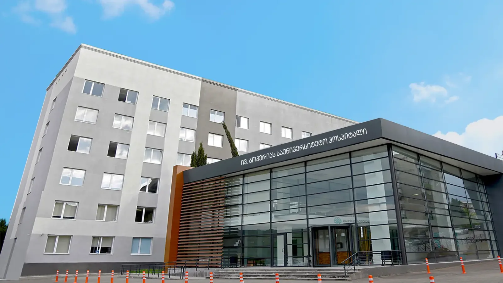
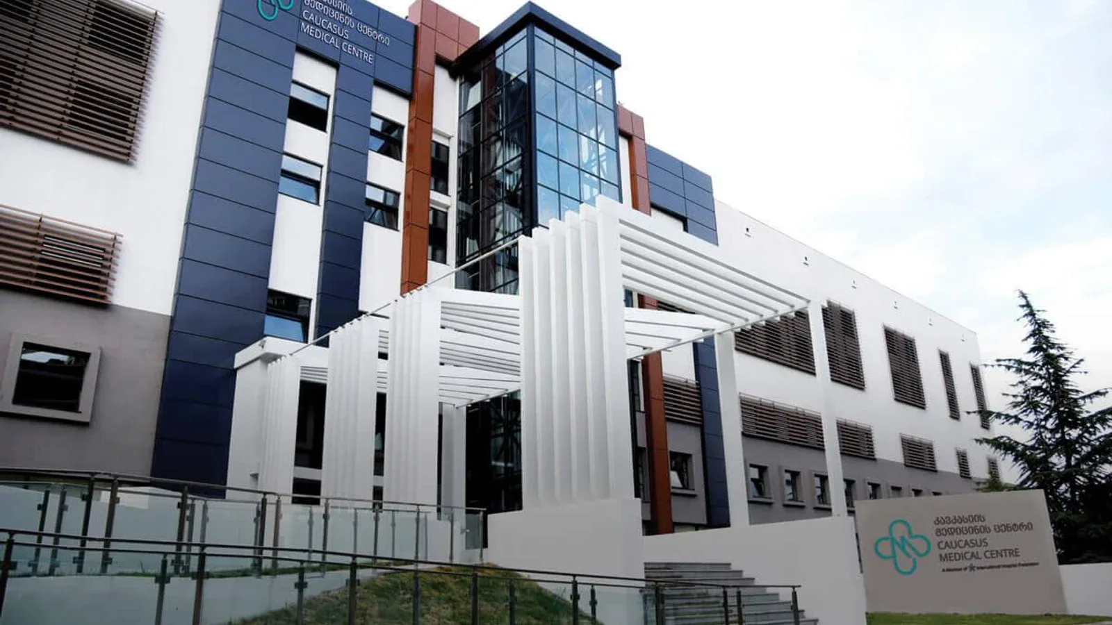
Cardiology and Cardiovascular Surgery care in Tbilisi is available at 3 hospitals in the Voumed network, with 3 related treatments.
Cardiology and cardiovascular surgery diagnose, treat and help prevent diseases of the heart and the blood vessels, from coronary artery disease and heart failure to rhythm disorders, valve disease and high blood pressure. Cardiology is the medical side, using examination, imaging and procedures performed through a thin catheter, while cardiovascular surgery operates on the heart and great vessels when an operation gives the best result. The two work as one team, so each patient follows a single, coherent plan. Heart disease is a leading cause of illness worldwide, yet much of it can be prevented, detected early or treated effectively, which is why timely, expert care matters so much. Patients often travel abroad for this care because it brings together experienced heart teams, advanced imaging and catheter laboratories, and shorter waiting times for planned treatment.
At a glance
- Sub-specialties
- clinical cardiology, interventional cardiology, electrophysiology, heart failure, cardiac imaging, cardiovascular surgery
- Common procedures
- coronary angiography and stenting, pacemaker and defibrillator implantation, ablation for rhythm disorders, coronary bypass, heart valve repair or replacement
- Common reasons to travel
- experienced heart teams, advanced catheter and imaging technology, shorter waiting times for planned surgery
- Typical hospital stay
- day case or one night for many catheter procedures, around 5 to 7 nights after open heart surgery
- Anaesthesia
- local with sedation for most catheter procedures, general anaesthesia for heart surgery
- Typical first step
- a consultation with an electrocardiogram and an echocardiogram, with further imaging or a catheter study if needed
Not sure which hospital fits your case?
Upload your medical records and let AI match you to the right hospital.
Upload records and get matchedAvailable at these hospitals






Procedures
Technologies and equipment
Photon-Counting CT
Photon-counting CT is a next-generation computed tomography technology with a new kind of detector. Where conventional CT detectors measure the total amount of X-ray energy that arrives, a photon-counting scanner counts each individual X-ray photon and measures its energy. This more refined way of gathering data produces noticeably sharper images with higher contrast, so very small structures, fine blood vessels and early-stage disease can be seen in greater detail, all while keeping the radiation dose low. Like all CT it uses X-rays, but it is designed to make the most of every photon, which can mean better images at a lower dose.
Hybrid Operating Room
A hybrid operating room is a surgical theatre that combines a full operating room with advanced, built-in medical imaging in the same space. Instead of relying on portable equipment or moving a patient to a separate scanning room, the team has a fixed, high-resolution imaging system, such as a robotic angiography arm, a CT scanner or an MRI, positioned right at the operating table. This lets surgeons see detailed live pictures of the body during the procedure and combine open surgery with minimally invasive, catheter-based techniques in a single session. For the patient, it can mean a less invasive operation, immediate confirmation that the surgery worked, fewer transfers between rooms and, often, a safer option when the case is complex or high-risk.
DSA Digital Subtraction Angiography
Digital subtraction angiography (DSA) is an advanced imaging method that shows the blood vessels throughout the body in fine detail. A thin catheter delivers a contrast agent into the arteries, and specialised computer processing strips away the surrounding bone and tissue so that only the vessels stand out sharply. It is used to detect vascular problems such as narrowing, aneurysm, malformation and abnormal connections in the brain, abdomen, skin and limbs. DSA is also the basis for many minimally invasive treatments, allowing a specialist to find and, in the same session, treat a vascular problem through a tiny entry point rather than open surgery.
Da Vinci Robotic Surgery
The da Vinci robotic surgical system lets a surgeon perform complex operations through a few small keyhole incisions instead of one large cut. Sitting at a nearby console, the surgeon controls tiny wristed instruments and a magnified high-definition three-dimensional camera, while the robotic arms translate every hand movement into precise, steady motion inside the body. The system never acts on its own: the surgeon is in full control at all times. For patients, this minimally invasive approach often means less pain, smaller scars, less blood loss and a quicker return to normal life.
Cardiac MRI
Cardiac MRI is a non-invasive imaging method that produces remarkably detailed pictures of the heart and the large blood vessels around it. It uses a strong magnetic field and radio waves rather than X-rays, so there is no ionising radiation involved. Unlike many other heart tests, it shows not only the shape and motion of the heart but also the condition of the heart muscle itself, down to the tissue level. This makes it a powerful tool for diagnosing and guiding the treatment of a wide range of heart conditions.
Coronary CT Angiography
Coronary CT angiography, sometimes called virtual angiography, is a computed tomography method that produces detailed images of the heart and its coronary arteries without the need for a catheter. Traditional angiography involves threading a thin tube into the arteries, but this scan does the same job from the outside, using a fast CT scanner and a contrast agent given through a vein. It clearly shows the build-up of calcium and plaque in the artery walls and any narrowing they cause. It uses X-rays, as all CT does, with techniques designed to keep the radiation dose low.
512-Slice CT
512-slice CT is a very fast, high-detail computed tomography scanner that captures a large number of thin image slices with each rotation. By gathering so much information so quickly, it can build detailed three-dimensional pictures of the body in a single short breath-hold. This speed is especially valuable for imaging moving organs such as the heart, where a fast scan freezes motion and produces sharp images. It uses X-rays, like all CT, but modern scanners of this kind are designed to keep the radiation dose as low as possible.
Dual-Energy CT
Dual-energy CT is an advanced form of computed tomography that scans the body at two different X-ray energy levels at the same time. A standard CT uses a single energy and shows mainly the shape and density of tissues, but by comparing how structures behave at two energies, dual-energy CT can tell different materials apart far more precisely. This added information helps doctors characterise what they see, such as distinguishing one type of tissue or deposit from another, and it can often be achieved with less contrast agent and a lower radiation dose. It uses X-rays, as all CT does, but with techniques designed to keep exposure low.
Not sure which hospital fits your case?
Upload your medical records and let AI match you to the right hospital.
Upload records and get matched