


Breast Health
Tbilisi

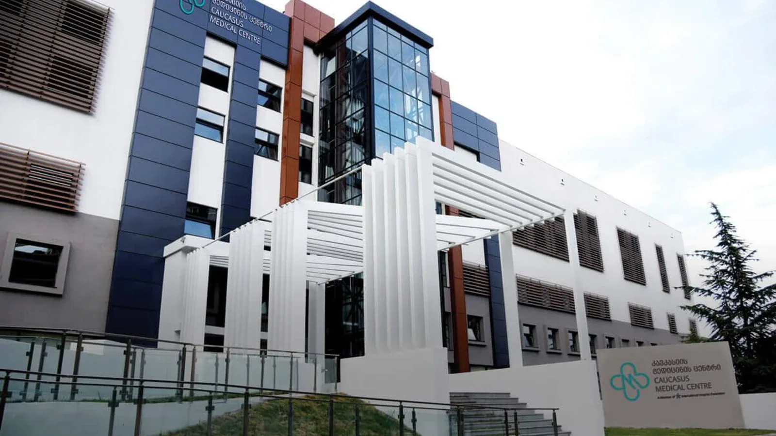
Breast Health care in Tbilisi is available at 1 hospital in the Voumed network.
Breast health care looks after the whole range of breast conditions, from harmless lumps and cysts to breast cancer, with one central aim: to find any serious disease early, when it is most treatable. Breast cancer is among the most common cancers in women, yet when it is caught while still small and confined to the breast, treatment is usually very successful. People travel abroad for breast care to reach experienced teams, modern imaging and the multidisciplinary approach that brings surgeons, oncologists, radiologists and pathologists together around a single plan. A dedicated breast service offers not only diagnosis and treatment but also reassurance, since most breast changes turn out to be benign.
At a glance
- Sub-specialties
- breast surgery, breast imaging and radiology, medical oncology, radiation oncology, pathology, oncoplastic and reconstructive surgery
- Common procedures
- clinical examination, mammography, breast ultrasound, breast MRI, needle biopsy, breast-conserving and reconstructive surgery
- Common reasons to travel
- experienced multidisciplinary teams, advanced imaging, integrated cancer care and the option of a second opinion
- Typical hospital stay
- outpatient for screening, diagnosis and biopsy, day case or 1 to 3 nights for breast surgery
- Anaesthesia
- local anaesthesia for most biopsies, general anaesthesia for breast surgery
- Typical first step
- a consultation with a clinical breast examination and imaging chosen for the individual
Not sure which hospital fits your case?
Upload your medical records and let AI match you to the right hospital.
Upload records and get matchedAvailable at these hospitals



Technologies and equipment
4D Breast Ultrasound
4D breast ultrasound, also known as automated breast volumetric scanning, is an imaging method that supports the diagnosis of breast cancer. Unlike a hand-held ultrasound, it uses a dedicated probe that moves automatically across the breast to capture the whole organ as a complete three-dimensional volume. It is mainly used alongside mammography, especially for women with dense breast tissue, where a denser background can hide lesions on a standard mammogram. The examination is comfortable, non-invasive and free of ionising radiation, making it a valuable additional layer in breast screening and assessment.
Intraoperative Radiotherapy (IORT)
Intraoperative radiotherapy, or IORT, is a way of giving a single, focused dose of radiation directly to the area at highest risk during the operation itself, right after the tumour has been removed and while the patient is still asleep under anaesthesia. With the surgical wound open, the team can place the radiation applicator exactly on the tissue that needs it and gently move skin and nearby organs out of the path of the beam. The result is treatment that is aimed precisely where cancer cells are most likely to remain, while healthy tissue is shielded. For suitable patients, especially in early breast cancer, one intraoperative dose can replace or greatly shorten several weeks of radiotherapy after surgery.
Digital Mammography
Digital mammography is a low-dose X-ray method used to screen for and detect breast cancer at an early stage. It captures very high-resolution digital images of the breast that a radiologist can examine and enhance on screen, revealing small nodules, masses and tiny specks of calcium that may not be felt or seen on other tests. Because it can find changes long before they cause symptoms, it is the cornerstone of breast cancer screening and one of the most effective tools for catching the disease when it is most treatable.
Tomosynthesis Mammography (3D Mammography)
Tomosynthesis mammography, often called 3D mammography, is an advanced form of digital mammography that builds a three-dimensional picture of the breast from a series of thin layers. Instead of a single flat image in which overlapping tissue can hide or mimic a problem, it lets the radiologist scroll through the breast slice by slice on a high-resolution screen. This makes small lesions and tumours easier to see and helps distinguish real findings from harmless overlapping tissue, which is especially valuable for screening and for women with dense breasts.
Not sure which hospital fits your case?
Upload your medical records and let AI match you to the right hospital.
Upload records and get matched