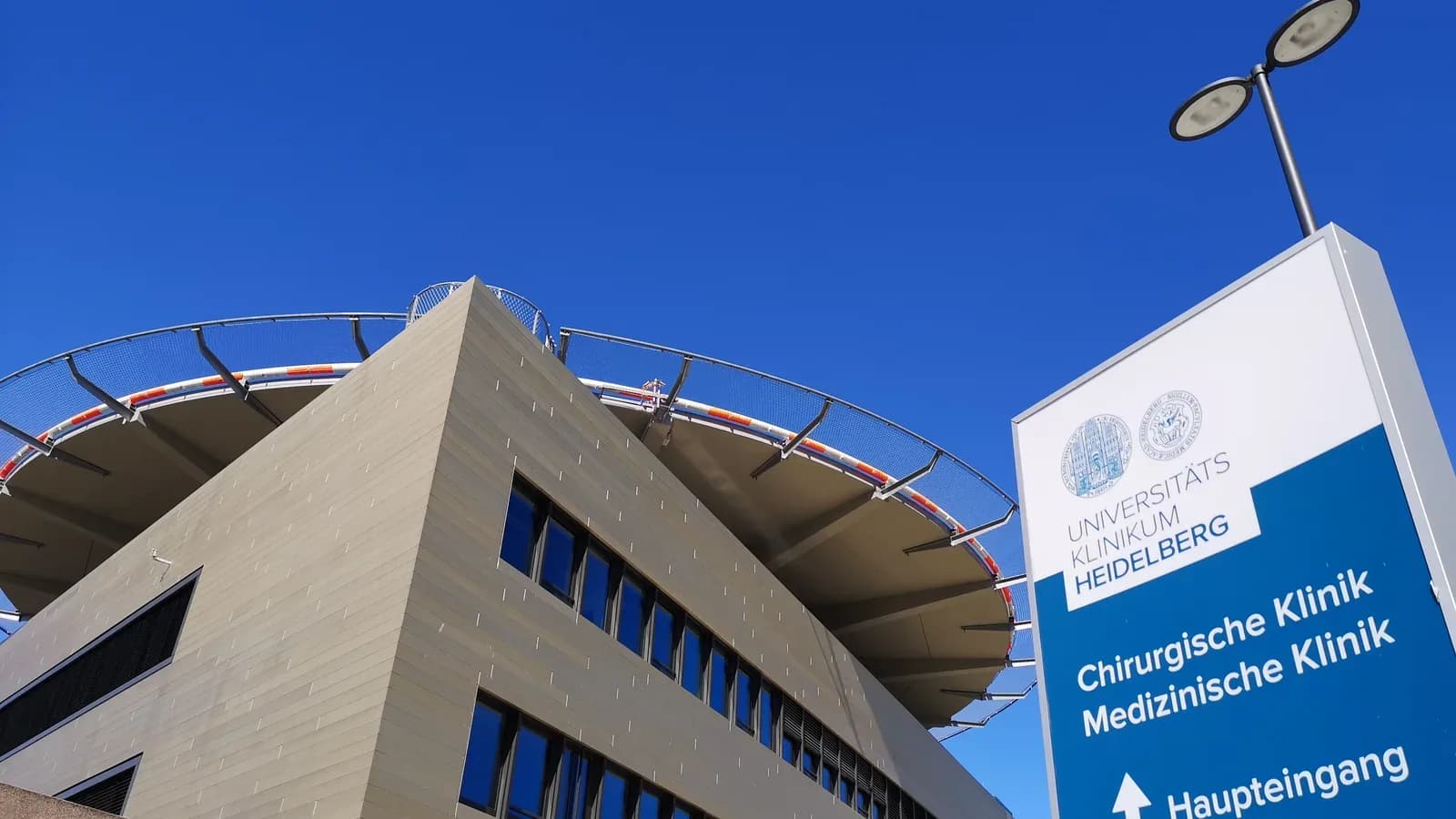
Heidelberg University Hospital
heidelberg
- Specialties
- 30
- Departments
- 37
Not sure which hospital fits your case?
Upload your medical records and let AI match you to the right hospital.
Upload records and get matchedAbout
Heidelberg University Hospital (Universitätsklinikum Heidelberg) is one of the leading academic medical centres in Germany and Europe, with a medical faculty dating back to 1818 and around 2500 beds. It is internationally renowned for cancer care as the home of the National Center for Tumor Diseases and the Heidelberg Ion-Beam Therapy Center, which delivers proton and carbon-ion radiotherapy, and it provides comprehensive multi-super-specialty care alongside leading research across oncology, cardiology, neuroscience, transplantation and nearly every other field of medicine.
Specialties
Departments
- Cardiac Sciences
- Cardiothoracic and Vascular Surgery
- Medical Oncology
- Radiation Oncology
- Surgical Oncology
- Breast Health
- Hematology and Bone Marrow Transplant
- Neurosciences
- Neurosurgery
- Spine Surgery
- Gastroenterology
- Gastrointestinal Surgery
- Orthopaedics and Joint Replacement
- Urology
- Nephrology
- Organ Transplantation
- Liver Transplant
- Kidney Transplant
- Pulmonology
- Thoracic Surgery
- Obstetrics and Gynaecology
- Reproductive Medicine and Fertility
- Paediatrics
- Neonatology
- Bariatric Surgery
- General Surgery
- Robotic Surgery
- Plastic and Reconstructive Surgery
- Dermatology
- Endocrinology and Diabetes Care
- Ophthalmology
- ENT
- Dentistry
- Rheumatology
- Sleep Medicine
- Physiotherapy and Rehabilitation
- Preventive Health and Check-up
Procedures
Bariatric and Metabolic Surgery
Cardiology and Cardiovascular Surgery
Dental, Oral and Maxillofacial Surgery
Ear, Nose and Throat
General Surgery
Gynecology and Obstetrics
IVF and Reproductive Health
Neurology and Neurosurgery
Ophthalmology
Organ Transplantation
Orthopedics and Traumatology
Physical Medicine and Rehabilitation
Plastic and Aesthetic Surgery
International patient services
- International patient office
- Interpreter and translation services
- Visa and travel assistance
- Airport transfer
- Accommodation assistance
Technologies and equipment
Carbon-Ion Therapy
Carbon-ion therapy is one of the most advanced forms of radiotherapy available, using a focused beam of carbon ions instead of conventional x-rays to destroy tumours with exceptional precision. Carbon ions are heavy charged particles that release most of their energy at a controlled depth inside the body, so the beam can be aimed to deliver its full dose inside the tumour while largely sparing the healthy tissue in front of it and beyond it. Because carbon ions also damage tumour cells more powerfully than standard radiation, this treatment is especially valuable for tumours that are difficult to remove surgically or that resist conventional radiotherapy. It is offered at only a small number of highly specialised particle-therapy centres worldwide.
CyberKnife M6
CyberKnife M6 is a robotic system for stereotactic radiosurgery and stereotactic body radiotherapy (SBRT). Despite the name, there is no knife and no cutting. A small linear accelerator sits on a computer-guided robotic arm and delivers many thin beams of focused radiation from hundreds of angles. The beams converge on the tumour with sub-millimetre accuracy, so a high dose reaches the target while nearby healthy tissue is spared. Imaging during treatment tracks the tumour continuously, and a motion-synchronisation feature follows targets that move with breathing, such as those in the lung or liver. Treatment is non-invasive and painless, needs no rigid head frame, and is usually given as an outpatient over one to five sessions. The decision is always made individually by the radiation oncology team.
PET-CT
PET-CT is an advanced hybrid imaging method that combines positron emission tomography with computed tomography in a single scan, mapping both the metabolic activity and the anatomical structure of the body at once. A small dose of a radioactive tracer, often a glucose analogue, is injected and gathers in cells that are working harder than normal, which is typical of many tumours. Because it can show where a disease is active before it changes the shape of an organ, PET-CT is one of the most valuable tools for detecting cancer, working out how far it has spread, and checking whether treatment is working.
Intraoperative Radiotherapy (IORT)
Intraoperative radiotherapy, or IORT, is a way of giving a single, focused dose of radiation directly to the area at highest risk during the operation itself, right after the tumour has been removed and while the patient is still asleep under anaesthesia. With the surgical wound open, the team can place the radiation applicator exactly on the tissue that needs it and gently move skin and nearby organs out of the path of the beam. The result is treatment that is aimed precisely where cancer cells are most likely to remain, while healthy tissue is shielded. For suitable patients, especially in early breast cancer, one intraoperative dose can replace or greatly shorten several weeks of radiotherapy after surgery.
Brachytherapy
Brachytherapy is a form of internal radiotherapy in which a radiation source is placed inside the body, right at or next to the tumour, rather than aimed from outside. Because the source sits so close to the target, it can deliver a high, very localised dose while the radiation falls off sharply over a short distance, sparing the healthy tissue around it. Modern systems automate this safely: thin tubes are guided to the tumour, the source travels through them along a precise plan, and it is withdrawn at the end, leaving no radiation behind in the body.
Da Vinci Robotic Surgery
The da Vinci robotic surgical system lets a surgeon perform complex operations through a few small keyhole incisions instead of one large cut. Sitting at a nearby console, the surgeon controls tiny wristed instruments and a magnified high-definition three-dimensional camera, while the robotic arms translate every hand movement into precise, steady motion inside the body. The system never acts on its own: the surgeon is in full control at all times. For patients, this minimally invasive approach often means less pain, smaller scars, less blood loss and a quicker return to normal life.
3 Tesla MRI
3 Tesla MRI is a high-field magnetic resonance imaging scanner that produces exceptionally detailed pictures of the inside of the body. The "3 Tesla" refers to the strength of its magnet, which is about twice that of a standard MRI scanner, and this extra power allows sharper, higher-resolution images, often in less time. Like all MRI, it uses a strong magnetic field and radio waves rather than X-rays, so there is no ionising radiation involved. It is especially valuable for examining the brain, the nervous system, joints and soft tissues, helping doctors detect and characterise problems that may be hard to see on other scans.
MR-Linac (MRI-Guided Radiotherapy)
MR-Linac combines a radiotherapy machine with an MRI scanner in a single device, so the radiation oncology team can watch the tumour and the soft tissue around it in real time while treatment is being delivered. Standard radiotherapy relies on planning scans taken on earlier days, but tumours and organs shift slightly from session to session and even with breathing. By seeing the target live, MR-Linac lets the team adapt the plan each day and pause or steer the beam as the tumour moves, delivering a precise dose where it is needed while better protecting healthy tissue.
Location
Im Neuenheimer Feld 672, 69120 Heidelberg, Germany
View on Google MapsNot sure which hospital fits your case?
Upload your medical records and let AI match you to the right hospital.
Upload records and get matched