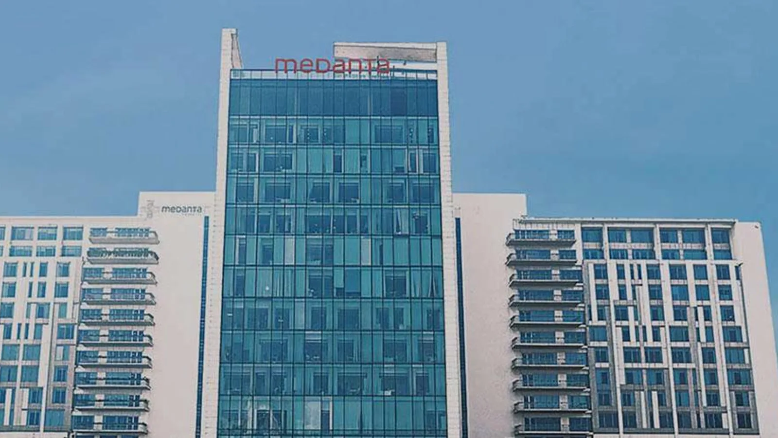
Medanta - The Medicity
gurugram
- Specialties
- 28
- Departments
- 35
Not sure which hospital fits your case?
Upload your medical records and let AI match you to the right hospital.
Upload records and get matchedAbout
Medanta - The Medicity, in Gurugram near New Delhi, is one of India's largest multi-super-specialty quaternary hospitals, founded in 2009 by the cardiac surgeon Dr. Naresh Trehan. The Gurugram campus has around 1250 beds and brings together institutes in cardiac sciences, cancer care, neurosciences, digestive and liver diseases, bone and joint care, kidney and urology and multi-organ transplantation. It is accredited by Joint Commission International (JCI) and by India's NABH and NABL, and is widely recognised for heart, liver, lung and kidney transplantation, bone-marrow transplantation, robotic surgery and advanced radiation oncology.
Specialties
Departments
- Cardiac Sciences
- Cardiothoracic and Vascular Surgery
- Medical Oncology
- Radiation Oncology
- Surgical Oncology
- Breast Health
- Hematology and Bone Marrow Transplant
- Neurosciences
- Neurosurgery
- Spine Surgery
- Gastroenterology
- Gastrointestinal Surgery
- Orthopaedics and Joint Replacement
- Urology
- Nephrology
- Organ Transplantation
- Liver Transplant
- Kidney Transplant
- Pulmonology
- Thoracic Surgery
- Obstetrics and Gynaecology
- Paediatrics
- Neonatology
- Bariatric Surgery
- General Surgery
- Robotic Surgery
- Plastic and Reconstructive Surgery
- Dermatology
- Endocrinology and Diabetes Care
- Ophthalmology
- ENT
- Dentistry
- Rheumatology
- Sleep Medicine
- Preventive Health and Check-up
Procedures
Bariatric and Metabolic Surgery
Cardiology and Cardiovascular Surgery
Dental, Oral and Maxillofacial Surgery
Ear, Nose and Throat
General Surgery
Gynecology and Obstetrics
Neurology and Neurosurgery
Organ Transplantation
Orthopedics and Traumatology
Plastic and Aesthetic Surgery
International patient services
- International patient office
- Interpreter and translation services
- Visa and travel assistance
- Airport transfer
- Accommodation assistance
Technologies and equipment
DSA Digital Subtraction Angiography
Digital subtraction angiography (DSA) is an advanced imaging method that shows the blood vessels throughout the body in fine detail. A thin catheter delivers a contrast agent into the arteries, and specialised computer processing strips away the surrounding bone and tissue so that only the vessels stand out sharply. It is used to detect vascular problems such as narrowing, aneurysm, malformation and abnormal connections in the brain, abdomen, skin and limbs. DSA is also the basis for many minimally invasive treatments, allowing a specialist to find and, in the same session, treat a vascular problem through a tiny entry point rather than open surgery.
Varian Edge
Varian Edge is an advanced radiosurgery platform, a high-precision machine that treats tumours with very focused beams of radiation delivered from outside the body, without any cutting. It is built for stereotactic radiosurgery and stereotactic body radiotherapy, two techniques that concentrate a high dose of radiation onto the exact shape of a target while protecting the healthy tissue around it. Image guidance and real-time motion management track tiny patient or tumour movement and keep the beam on target with sub-millimetre accuracy. Treatment is painless, needs no rigid head frame for brain targets, and is usually completed in a small number of short outpatient sessions. The radiation oncology team always plans each case individually.
TomoTherapy
TomoTherapy is an image-guided radiotherapy system that delivers radiation in a continuous spiral as the treatment ring rotates around the patient, much like a CT scanner. Built-in CT imaging lets the radiation oncology team confirm the tumour's exact position before every session, and the beam is divided into many small beamlets that paint the dose precisely onto the target. This slice-by-slice approach is well suited to complex or unusually shaped tumours and to large or long treatment areas, while keeping nearby healthy organs better protected.
CyberKnife M6
CyberKnife M6 is a robotic system for stereotactic radiosurgery and stereotactic body radiotherapy (SBRT). Despite the name, there is no knife and no cutting. A small linear accelerator sits on a computer-guided robotic arm and delivers many thin beams of focused radiation from hundreds of angles. The beams converge on the tumour with sub-millimetre accuracy, so a high dose reaches the target while nearby healthy tissue is spared. Imaging during treatment tracks the tumour continuously, and a motion-synchronisation feature follows targets that move with breathing, such as those in the lung or liver. Treatment is non-invasive and painless, needs no rigid head frame, and is usually given as an outpatient over one to five sessions. The decision is always made individually by the radiation oncology team.
TrueBeam STx
TrueBeam STx is an advanced linear accelerator, a machine that delivers external radiotherapy to treat cancer with very high precision. It shapes powerful radiation beams to match the exact size and shape of a tumour and aims them from many angles, so that a strong dose reaches the target while nearby healthy tissue and organs receive as little as possible. Because it tracks the target and can account for movement such as breathing, it is accurate to within millimetres. This makes it suitable both for conventional, daily radiotherapy and for advanced focused techniques that treat a tumour in only a few sessions. The treatment is non-invasive and painless, with nothing entering the body.
Da Vinci Robotic Surgery
The da Vinci robotic surgical system lets a surgeon perform complex operations through a few small keyhole incisions instead of one large cut. Sitting at a nearby console, the surgeon controls tiny wristed instruments and a magnified high-definition three-dimensional camera, while the robotic arms translate every hand movement into precise, steady motion inside the body. The system never acts on its own: the surgeon is in full control at all times. For patients, this minimally invasive approach often means less pain, smaller scars, less blood loss and a quicker return to normal life.
O-Arm
The O-arm is an intraoperative imaging system that rotates a full circle around the patient to produce real-time, high-resolution cross-sectional images while surgery is underway. In effect it brings a mobile CT-style scanner into the operating room, so the surgeon can see the exact position of bone, instruments and implants at the moment they are being placed, rather than relying only on images taken before the operation. It is used mainly in spine, brain and nerve, and orthopaedic trauma surgery, where it gives precise guidance for critical steps and supports greater accuracy and safety.
3 Tesla MRI
3 Tesla MRI is a high-field magnetic resonance imaging scanner that produces exceptionally detailed pictures of the inside of the body. The "3 Tesla" refers to the strength of its magnet, which is about twice that of a standard MRI scanner, and this extra power allows sharper, higher-resolution images, often in less time. Like all MRI, it uses a strong magnetic field and radio waves rather than X-rays, so there is no ionising radiation involved. It is especially valuable for examining the brain, the nervous system, joints and soft tissues, helping doctors detect and characterise problems that may be hard to see on other scans.
Intraoperative MRI
Intraoperative MRI, also called operating room MRI, brings the power of magnetic resonance imaging directly into surgery. A specially designed scanner, integrated into the operating room, lets the surgical team obtain detailed pictures of the brain or spine while the operation is still under way. This means the surgeon can check progress during the procedure rather than waiting for a scan afterwards. Like all MRI, it uses a magnetic field and radio waves instead of X-rays, so it adds no ionising radiation. It is especially valuable for delicate tumour surgery, where seeing the result in real time can improve the outcome.
Location
CH Baktawar Singh Road, Sector 38, Gurugram, Haryana 122001, India
View on Google MapsAccreditations
- JCI
- NABH
Not sure which hospital fits your case?
Upload your medical records and let AI match you to the right hospital.
Upload records and get matched