

Mediclinic City Hospital
Dubai

- Specialties
- 28
- Departments
- 41
Not sure which hospital fits your case?
Upload your medical records and let AI match you to the right hospital.
Upload records and get matchedAbout
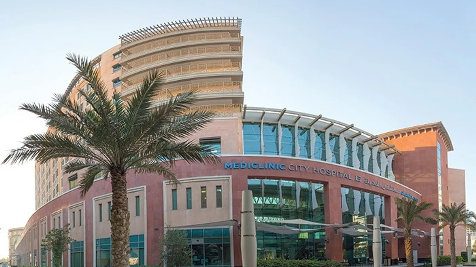
Mediclinic City Hospital is a 280 bed private hospital in Dubai Healthcare City and part of the Mediclinic Middle East network. Alongside an extensive outpatient department it operates 27 neonatal beds, 27 intensive care beds, six operating rooms, three daycare surgery units, a caesarean section theatre, two cardiac catheterisation laboratories, endoscopy suites, an on site laboratory, an emergency department, labour and post natal wards, a 24 hour pharmacy and a radiology department. Dedicated units cover oncology, breast disease, cardiothoracic surgery, neonatal and paediatric intensive care and stroke medicine. The stroke unit holds accreditation from the German Stroke Society, the hospital carries ASA/AHA International Certification as a Comprehensive Stroke Center, and its Acute Coronary Syndrome programme is certified by Joint Commission International. The North Wing, opened in 2016, houses the Comprehensive Cancer Centre together with a daycare surgery ward, endoscopy suites, an extended dialysis unit, rehabilitation physiotherapy, orthotics and prosthetics services and a reference laboratory. The transplant centre performs living donor and deceased donor kidney transplants, and the hospital carried out the first kidney transplant in Dubai in 2016 and the first robotic assisted knee replacement in the Middle East in 2018. Technology in use includes PET/CT, SPECT CT, 3T MRI, digital mammography, a digital bone densitometer and a digital catheterisation laboratory, and surgical services use the da Vinci Xi HD four arm robotic system. The hospital is accredited by Joint Commission International.
Specialties
Physicians
1-12 of 139
Departments
- Cardiology
- Cardiothoracic Surgery
- Comprehensive Cancer Centre
- Nuclear Medicine
- Haematology
- Breast Centre
- Neurology
- Neurosurgery
- Stroke Unit
- Spine Surgery
- Orthopaedics
- Sports and Exercise Medicine
- Robotic Surgery
- General Surgery
- Hernia Surgery
- Gastrointestinal Surgery
- Gastroenterology
- Liver Clinic
- Bariatric Surgery
- Obesity Management Programme
- Urology
- Kidney Care
- Transplant Centre
- Obstetrics and Gynaecology
- Fetal Medicine
- Fertility
- Paediatrics
- Neonatal Intensive Care Unit
- Paediatric Intensive Care Unit
- Ophthalmology
- Ear, Nose and Throat
- Dermatology
- Endocrinology and Diabetes
- Dentistry
- Orthodontics
- Plastic, Reconstructive and Aesthetic Surgery
- Pulmonology and Respiratory Diseases
- Rheumatology
- Physiotherapy and Rehabilitation
- Orthotics and Prosthetics
- Pain Medicine
Procedures
Bariatric and Metabolic Surgery
Cardiology and Cardiovascular Surgery
Dental, Oral and Maxillofacial Surgery
Ear, Nose and Throat
Gastroenterology
General Surgery
Gynecology and Obstetrics
IVF and Reproductive Health
Ophthalmology
Organ Transplantation
Orthopedics and Traumatology
Physical Medicine and Rehabilitation
Plastic and Aesthetic Surgery
Urology
International patient services
- Cross border telemedicine consultations
- Airport meet and greet service
- Short and long term hotel reservations
- Language translation services
- Inbound visa support for the United Arab Emirates
- Airport transfer and chauffeur services
Technologies and equipment
EBUS Endobronchial Ultrasound
Endobronchial ultrasound (EBUS) is a minimally invasive method that combines bronchoscopy with ultrasound to examine the airways and the tissues and lymph nodes that surround them. A thin, flexible bronchoscope carrying a camera and a small ultrasound probe is passed through the mouth into the airways, where the probe creates real-time images of structures just beyond the airway wall, such as lymph nodes and masses that cannot be seen with a camera alone. Under this live ultrasound guidance, a fine needle can take samples for the laboratory in the same session, all without any surgical incision.
PET-CT
PET-CT is an advanced hybrid imaging method that combines positron emission tomography with computed tomography in a single scan, mapping both the metabolic activity and the anatomical structure of the body at once. A small dose of a radioactive tracer, often a glucose analogue, is injected and gathers in cells that are working harder than normal, which is typical of many tumours. Because it can show where a disease is active before it changes the shape of an organ, PET-CT is one of the most valuable tools for detecting cancer, working out how far it has spread, and checking whether treatment is working.
FibroScan
FibroScan is a non-invasive, ultrasound-based device that measures how stiff the liver is, which reflects the degree of scarring, or fibrosis, and at the same time estimates the amount of fat in the liver. It offers a fast, painless alternative to a liver biopsy, with no incision or needle, and it assesses a larger area of the organ than a tiny tissue sample would. A probe is simply placed on the skin over the liver while the patient lies down, and a numerical result is available within minutes. It is widely used to detect and follow liver conditions and to guide and monitor treatment.
SPECT-CT
SPECT-CT is a nuclear medicine imaging method that merges single photon emission computed tomography with computed tomography in one device, capturing both how an organ functions and its anatomical structure in a single session. A low-dose radiopharmaceutical is injected and gathers in the target tissue, where a rotating gamma camera builds three-dimensional functional images while the CT scan defines the exact location within the body. By showing not just the shape of a structure but how active it is, SPECT-CT helps doctors find disease, pinpoint exactly where it sits, and plan treatment with greater confidence.
Brachytherapy
Brachytherapy is a form of internal radiotherapy in which a radiation source is placed inside the body, right at or next to the tumour, rather than aimed from outside. Because the source sits so close to the target, it can deliver a high, very localised dose while the radiation falls off sharply over a short distance, sparing the healthy tissue around it. Modern systems automate this safely: thin tubes are guided to the tumour, the source travels through them along a precise plan, and it is withdrawn at the end, leaving no radiation behind in the body.
DSA Digital Subtraction Angiography
Digital subtraction angiography (DSA) is an advanced imaging method that shows the blood vessels throughout the body in fine detail. A thin catheter delivers a contrast agent into the arteries, and specialised computer processing strips away the surrounding bone and tissue so that only the vessels stand out sharply. It is used to detect vascular problems such as narrowing, aneurysm, malformation and abnormal connections in the brain, abdomen, skin and limbs. DSA is also the basis for many minimally invasive treatments, allowing a specialist to find and, in the same session, treat a vascular problem through a tiny entry point rather than open surgery.
ESWT (Extracorporeal Shock Wave Therapy)
Extracorporeal shock wave therapy (ESWT) is a non-surgical, drug-free treatment that delivers high-energy acoustic waves generated outside the body to a painful or slow-healing tissue. The waves pass harmlessly through the skin and concentrate on the target area, where they stimulate the body's own repair processes. ESWT is used mainly for long-standing tendon, joint and soft-tissue problems that have not responded well to rest, medication or standard physiotherapy. Sessions are short, done in the clinic without anaesthesia, and afterwards most people return straight to their day. It is delivered as part of a rehabilitation plan rather than on its own.
ECMO
ECMO, short for extracorporeal membrane oxygenation, is an advanced life-support technology that does the work of the heart, the lungs, or both, when they are too sick to keep the body supplied with oxygen. Blood is drawn out of the body through a tube, passed through a machine that adds oxygen and removes carbon dioxide just as healthy lungs would, and then returned warmed to the body. By taking over this vital task, ECMO gives badly damaged organs a chance to rest and recover, or it keeps a patient alive while the medical team treats the underlying cause or arranges further treatment. It is used only in intensive care for the most serious, but potentially reversible, heart and lung emergencies, and it is run continuously by a specialised team around the clock. Because it demands rare expertise and resources, ECMO is available only in a small number of highly equipped centres.
Da Vinci Robotic Surgery
The da Vinci robotic surgical system lets a surgeon perform complex operations through a few small keyhole incisions instead of one large cut. Sitting at a nearby console, the surgeon controls tiny wristed instruments and a magnified high-definition three-dimensional camera, while the robotic arms translate every hand movement into precise, steady motion inside the body. The system never acts on its own: the surgeon is in full control at all times. For patients, this minimally invasive approach often means less pain, smaller scars, less blood loss and a quicker return to normal life.
O-Arm
The O-arm is an intraoperative imaging system that rotates a full circle around the patient to produce real-time, high-resolution cross-sectional images while surgery is underway. In effect it brings a mobile CT-style scanner into the operating room, so the surgeon can see the exact position of bone, instruments and implants at the moment they are being placed, rather than relying only on images taken before the operation. It is used mainly in spine, brain and nerve, and orthopaedic trauma surgery, where it gives precise guidance for critical steps and supports greater accuracy and safety.
3 Tesla MRI
3 Tesla MRI is a high-field magnetic resonance imaging scanner that produces exceptionally detailed pictures of the inside of the body. The "3 Tesla" refers to the strength of its magnet, which is about twice that of a standard MRI scanner, and this extra power allows sharper, higher-resolution images, often in less time. Like all MRI, it uses a strong magnetic field and radio waves rather than X-rays, so there is no ionising radiation involved. It is especially valuable for examining the brain, the nervous system, joints and soft tissues, helping doctors detect and characterise problems that may be hard to see on other scans.
Cardiac MRI
Cardiac MRI is a non-invasive imaging method that produces remarkably detailed pictures of the heart and the large blood vessels around it. It uses a strong magnetic field and radio waves rather than X-rays, so there is no ionising radiation involved. Unlike many other heart tests, it shows not only the shape and motion of the heart but also the condition of the heart muscle itself, down to the tissue level. This makes it a powerful tool for diagnosing and guiding the treatment of a wide range of heart conditions.
Digital Mammography
Digital mammography is a low-dose X-ray method used to screen for and detect breast cancer at an early stage. It captures very high-resolution digital images of the breast that a radiologist can examine and enhance on screen, revealing small nodules, masses and tiny specks of calcium that may not be felt or seen on other tests. Because it can find changes long before they cause symptoms, it is the cornerstone of breast cancer screening and one of the most effective tools for catching the disease when it is most treatable.
Tomosynthesis Mammography (3D Mammography)
Tomosynthesis mammography, often called 3D mammography, is an advanced form of digital mammography that builds a three-dimensional picture of the breast from a series of thin layers. Instead of a single flat image in which overlapping tissue can hide or mimic a problem, it lets the radiologist scroll through the breast slice by slice on a high-resolution screen. This makes small lesions and tumours easier to see and helps distinguish real findings from harmless overlapping tissue, which is especially valuable for screening and for women with dense breasts.
Endoscopic Ultrasound (EUS)
Endoscopic ultrasound (EUS) combines endoscopy and ultrasound in a single thin instrument, allowing the deeper layers of the digestive tract and the organs and tissues around it to be examined in detail. By placing a tiny ultrasound probe at the tip of an endoscope and guiding it inside the body, very close to the area of interest, it produces highly detailed images of structures such as the pancreas, bile ducts and nearby lymph nodes that can be hard to see from the outside. When needed, a fine needle can take a sample for the laboratory during the same procedure, all without any surgical incision.
Robotic Arm-Assisted Orthopedic Surgery
Robotic arm-assisted orthopedic surgery is a technology used mainly in knee and hip replacement to plan and carry out the operation with very high accuracy. A detailed three-dimensional plan is built from the patient's own CT scan, and during surgery a robotic arm guides the surgeon's instruments so that bone is prepared and the implant is positioned to that exact plan. The surgeon always holds and directs the instrument; the robotic arm adds steadiness and built-in limits that protect the surrounding tissue. The aim is a joint that fits and balances well, which can mean less pain and a smoother recovery.
Rezum (Water Vapor Therapy)
Rezum is a minimally invasive treatment for an enlarged prostate that uses the natural energy stored in water vapour, or steam, to gently shrink the overgrown tissue. Benign prostatic enlargement is a common condition in older men, where the prostate grows and presses on the urinary channel, causing symptoms such as a weak stream, frequent urination and getting up at night. Rezum delivers small, precise bursts of steam directly into the enlarged tissue through the urethra, with no cutting. Over the following weeks the treated tissue is naturally reabsorbed, the channel opens up, and urinary symptoms improve.
Neuronavigation
Neuronavigation is an image-guided surgical system that acts like a precise map and GPS for the brain and spine. Using the patient's own scans built into a three-dimensional model, it shows the surgeon exactly where the instruments are inside the body in real time, so the safest, shortest route to a lesion can be planned and followed. This is especially important in the brain and spine, where targets are often small, deep and surrounded by critical nerves and blood vessels. By guiding the surgeon away from healthy structures, neuronavigation supports accuracy, smaller approaches and added safety.
pH-metry (Reflux Diagnosis)
pH-metry is a diagnostic test that confirms reflux disease by directly measuring how much acid reaches the food pipe (oesophagus) over an extended period. It is especially useful when standard endoscopy looks normal but a person still has reflux-type symptoms, because it captures acid exposure that a single snapshot examination would miss. In a common modern version, a tiny pH capsule is attached to the lower oesophagus during a brief endoscopy under sedation, then records acid levels for about 24 to 48 hours before detaching on its own and passing naturally. The result gives an objective picture of whether, and how often, acid is reaching the oesophagus.
Location
Mediclinic City Hospital, Dubai Healthcare City, P O Box 505004, Dubai, United Arab Emirates
View on Google MapsAccreditations
- JCI
Not sure which hospital fits your case?
Upload your medical records and let AI match you to the right hospital.
Upload records and get matched