Jack Campbell Injury: Protocols, Hospital Assessments, and Recovery After Sports Cervical Trauma
Jack Campbell injury protocols examine on-field spinal immobilization, emergency CT and MRI scans, and recovery pathways for sports cervical trauma.

Quick Answer: On-field backboarding and hospital evaluation after a high-impact collision are standard precautionary protocols. They immobilize the cervical spine and protect neural pathways while emergency physicians conduct definitive diagnostic imaging to rule out structural instability.
Key Takeaways:
- Spinal immobilization with a backboard and rigid collar prevents spinal cord displacement following blunt impact until imaging confirms structural stability.
- A stinger involves a transient peripheral nerve stretch affecting one arm, whereas structural cervical trauma causes midline bony tenderness or bilateral symptoms.
- Non-contrast computed tomography (CT) serves as the primary emergency scan for bone fractures, while magnetic resonance imaging (MRI) evaluates soft tissues, discs, and the spinal cord.
- Non-operative care, including brief rest, structured rehabilitation, and progressive isometric strengthening, resolves most stable sports-related neck conditions.
- Immediate emergency care is necessary if neck trauma causes bilateral numbness, midline spinal tenderness, progressive motor weakness, or changes in bladder function.
Cervical spine trauma in collision sports spans a wide spectrum, from benign muscle strains and peripheral nerve stretches to disc herniations, vertebral fractures, and spinal cord injuries. Because early muscle spasms can mimic unstable fractures, clinical protocols require medical staff to treat severe impacts as potentially unstable until diagnostic imaging confirms otherwise.
Video · 2:27
Jack Campbell Injury Explained: Why a Backboard, and What Happens Next
Video transcript
0:00 IntroJack Campbell was carted off on a backboard after a hard tackle. What does that mean, and what happens next? Here is how medical teams and hospitals handle a possible neck injury, step by step.
0:11 What happenedOn November first against the Minnesota Vikings, Campbell went down after a tackle in the fourth quarter. He was carted off, taken to hospital for a neck evaluation, and kept overnight for continued monitoring. The team has not shared a diagnosis and says it will give updates as it has them.
0:27 Why a backboardA backboard and a collar are a precaution. They keep the head and neck still, so an injury that might be unstable cannot get worse on the way to hospital. They stay on until scans rule it out.
0:38 The neckThe neck, or cervical spine, has seven vertebrae from C1 to C7. Together with the discs and ligaments between them, they surround and protect the spinal cord, which is why any possible damage is taken seriously.
0:51 On the fieldOn the field, the medical team checks breathing, movement, and feeling, while someone holds the head in line. Helmet and shoulder pads usually stay on together, because removing only one of them can bend the neck.
1:03 Stinger or spinal cordNot every hit is a spine injury. A stinger, a stretched or pinched nerve, causes burning or tingling down one arm and usually settles within a day, though some weakness can last several days. Symptoms in both arms or in the legs point to the spinal cord.
1:18 In hospitalIn hospital, doctors first test strength, feeling, and reflexes in the arms and legs. Decision rules, such as the Canadian C-Spine rule and NEXUS, help them decide who needs a scan.
1:29 ScansA CT scan usually comes first to check the bones. An MRI shows the cord, discs, and ligaments if there are nerve symptoms. The collar comes off once the spine is cleared.
1:40 TreatmentMost stable neck injuries, like strains and stingers, are treated without surgery, with a short rest and then gentle movement. Surgery is for an unstable spine, or when the spinal cord or nerves are being compressed.
1:53 RecoveryNeck pain from a strain usually lasts a few weeks, and full recovery can take up to about eight weeks. A return to contact sport needs no pain, full movement, full strength, and a normal nerve exam.
2:06 Warning signsAfter any neck injury, call emergency services, do not move the person and do not remove the helmet if there is numbness or tingling in an arm or leg, weakness, loss of bladder or bowel control, or confusion. For the full guide with sources, visit the Voumed blog. And if this helped, subscribe for clear, sourced health explainers.
Jack Campbell Injury: Understanding On-Field Spinal Protocols
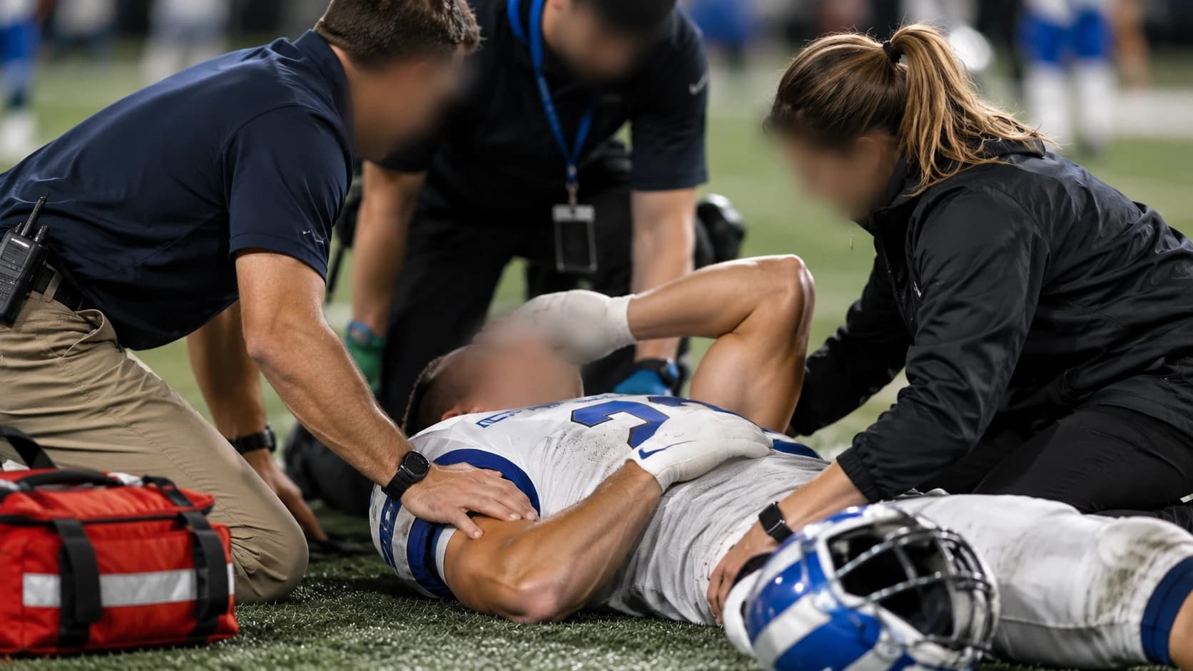
Detroit Lions linebacker Jack Campbell exited the November 1, 2026 game against the Minnesota Vikings on a backboard following a collision, remaining hospitalized overnight for neurological observation. On-field spinal immobilization serves as a standard precautionary procedure rather than proof of structural cord damage. It protects the cervical spine until emergency hospital scans exclude spinal instability.
During high-speed defensive collisions in professional football, the head and neck can undergo sudden flexion, hyperextension, or axial loading. In Campbell's case, medical staff initiated emergency protocols immediately after the hit, securing him with a rigid spine board, applying lateral head blocks, and transporting him off the field. Observers often view backboards as a sign of catastrophic injury; clinically, however, sports medicine physicians use spinal immobilization as a standard risk-mitigation step. Medical personnel operate under a clear directive: any athlete reporting neck discomfort or showing altered biomechanics after a collision must be protected against secondary movement.
Following transfer to an emergency trauma center, overnight hospital observation allows clinical teams to perform serial neurological evaluations. These repeated assessments ensure that delayed swelling, epidural hematomas, or subtle neurological deficits do not emerge as acute adrenaline subsides. Without an official diagnostic disclosure, medical commentary focuses on standard clinical algorithms rather than speculation about specific tissue damage.
Why Emergency Staff Use Backboards and Rigid Collars for a Neck Injury in Football
On-field athletic trainers and emergency medical staff apply rigid cervical collars and backboards immediately after suspected neck trauma to maintain neutral spinal alignment and prevent secondary neurologic deficits. Any uncontrolled movement of an unstable cervical vertebra could compress the spinal cord or adjacent nerve roots, making complete physical stabilization critical before imaging.
The cervical spine consists of seven small, highly mobile vertebrae (C1 through C7) that encase the upper spinal cord. When an impact generates sufficient force to cause a suspected neck injury in football, micro-motion from an unstable bone fragment or torn ligament can lead to spinal cord compression. To prevent this, athletic trainers, team physicians, and paramedics adhere to structured on-field protocols:
- Primary Airway and Neurological Triage: Clinicians simultaneously assess airway patency, spontaneous breathing, and responsiveness while maintaining manual in-line cervical stabilization.
- Log-Roll or Lift Maneuvers: If the player is prone or awkwardly positioned, a trained multi-person team executes a coordinated log-roll or lift-and-slide maneuver, keeping the head, shoulders, and pelvis aligned along a single axis.
- Equipment Management: Current sports trauma guidelines dictate that helmets and shoulder pads generally remain in place together unless the airway is compromised. Removing a helmet while leaving shoulder pads flexes the cervical spine forward, whereas leaving both in place preserves neutral spinal alignment.
- Multi-Point Fixation: Straps secure the torso, pelvis, and legs to the board, while head blocks and tape anchor the helmet to prevent lateral rotation during ambulance transit.
This controlled extraction minimizes mechanical movement during transit, helping prevent any occult mechanical instability from worsening before emergency department evaluation.
Stinger Injury Neck vs. Structural Trauma: Sorting Types of Cervical Impact
A stinger injury neck, often called a burner, occurs when brachial plexus nerves stretch or compress during a shoulder tackle, causing acute, temporary burning numbness down one arm. In contrast, structural trauma involves cervical muscle strains, herniated intervertebral discs, or cervical spine fractures that present with bilateral symptoms, central midline bony pain, or motor loss.
A stinger develops through two main mechanisms: lateral flexion of the neck away from the impacted shoulder (traction on the brachial plexus), or hyperextension with neck compression toward the affected side (neural foramen pinch). Patients typically describe an immediate, electric, burning sensation radiating from the neck into the shoulder, forearm, and fingers. Symptoms are strictly unilateral and typically resolve within seconds to several minutes, though deltoid or biceps weakness can occasionally persist for several days.
By contrast, a neck strain injury involves micro-tears in paraspinal muscles (such as the splenius capitis or trapezius) without direct nerve root impingement. Strains produce stiffness, local muscle spasms, and reduced active range of motion, but they do not cause electric pain radiating down the arm. Structural trauma, such as cervical disc tears or vertebral bone fractures, presents a far higher clinical risk. When an intervertebral disc herniates acutely or vertebral margins fracture under axial load, neural tissue may suffer direct compression, producing bilateral numbness, progressive weakness, or spinal shock.
| Cervical Injury Category | Primary Mechanism | Characteristic Symptoms | Primary Diagnostic Tool | Standard Management Approach |
|---|---|---|---|---|
| Stinger / Burner | Traction or compression of brachial plexus | Unilateral burning pain, arm weakness, transient numbness | Clinical exam; MRI if persistent | Relative rest, rotator cuff and neck strengthening, collar pads |
| Cervical Muscle Strain | Hyperflexion, hyperextension, or violent rotation | Paraspinal tightness, muscular spasm, localized ache | Clinical evaluation, X-ray to rule out bone injury | Soft tissue therapy, gentle active mobility, oral anti-inflammatories |
| Disc Herniation / Radiculopathy | Axial compression with rotation or flexion | Unilateral or bilateral shooting nerve pain, dermatomal numbness | Cervical MRI | Physical therapy, selective epidural injections, occasional discectomy |
| Cervical Vertebral Fracture | Direct axial loading (spearing or crown-of-helmet impact) | Midline bony tenderness, severe spasm, neurological deficits | Non-contrast CT scan | Rigid external orthosis (halo/cervical collar) or surgical stabilization |
| Cervical Cord Neuropraxia | Transient spinal cord compression in a narrowed canal | Bilateral numbness, tingling, or temporary motor weakness in all limbs | Cervical CT and MRI | Strict immobilization, immediate trauma evaluation, canal diameter analysis |
In-Hospital Assessment: CT, MRI, and Neurological Examinations for Neck Vertebrae Injury
Hospital evaluations for a suspected neck vertebrae injury prioritize a non-contrast cervical computed tomography (CT) scan to check for bone fractures and alignment disruption. If imaging clears bony trauma but neurological symptoms or spinal tenderness persist, clinicians perform magnetic resonance imaging (MRI) to inspect the spinal cord, intervertebral discs, and posterior ligamentous complex.
The trauma team follows a standardized clinical pathway upon receiving an immobilized athlete:
- Physical and Neurological Evaluation: The examining physician evaluates cranial nerves, motor strength across standard myotomes (C5 shoulder abduction through T1 finger abduction), sensory dermatomes, and deep tendon reflexes. Palpation of the posterior midline determines whether tenderness involves lateral soft tissues or lies directly over bony spinous processes.
- Computed Tomography (CT): A thin-slice, non-contrast CT scan of the cervical spine is the initial standard for detecting cortical bone fractures, facet dislocations, and canal compromise. CT scans take only minutes, rapidly identifying whether surgical stabilization is necessary.
- Magnetic Resonance Imaging (MRI): While CT captures bone detail, MRI evaluates soft tissues. It identifies spinal cord contusions (edema or hemorrhage within neural tissue), acute disc extrusions, and injuries to the posterior longitudinal ligament or ligamentum flavum. An intact ligamentous complex is critical for confirming that a cervical segment is mechanically stable.
- Serial Observation: Clinicians repeat neurological testing at scheduled intervals. If imaging confirms spinal stability and symptoms resolve, physicians transition the patient from rigid immobilization to a soft collar or active observation.
Clinical Pathways: Conservative Care to Surgical Intervention in Neck Injury Treatment
Modern neck injury treatment relies on non-operative measures for stable strains, stingers, and non-displaced injuries, using brief rest, physical therapy, nonsteroidal anti-inflammatory drugs, and isometric strengthening. Decompressive surgery or spinal fusion is reserved for progressive neurologic deficits, cervical fracture dislocations, or documented mechanical instability.
When imaging confirms that the spine is structurally intact, conservative rehabilitation proceeds through distinct phases:
- Acute De-loading (Days 1 to 5): The primary goal is reducing muscle spasms and protecting irritated nerves. Management includes short-term activity reduction, cryotherapy, and short courses of nonsteroidal anti-inflammatory medications. Prolonged rigid immobilization is avoided when instability is absent, as early guided motion helps prevent muscle stiffness.
- Active Range of Motion and Isometric Restoration (Weeks 1 to 3): Physical therapists introduce gentle active-assisted neck movements, scapular stabilization drills, and sub-maximal isometric contractions in neutral postures.
- Progressive Strengthening and Dynamic Stabilization (Weeks 3 to 6+): Rehabilitation advances to dynamic resistance training for cervical extensors, deep neck flexors, trapezius, and deltoids. Core control and kinetic-chain coordination are integrated to help the spine absorb future impact forces effectively.
Surgical intervention becomes necessary only under specific structural conditions: progressive motor weakness, acute disc herniation causing severe cord impingement, or high-grade facet subluxation. Surgical procedures, such as anterior cervical discectomy and fusion (ACDF) or posterior spinal instrumentation, aim to decompress neural structures and re-establish biomechanical stability.
Athletes must satisfy strict objective criteria before returning to contact sports: complete absence of neck pain, restoration of full active range of motion, return to baseline strength across all upper extremity muscle groups, and normal repeat clinical examinations.
Neck Injury When to Worry: Red Flag Signs After Sports, Falls, and Car Crashes
Critical indicators that demand immediate emergency intervention after neck trauma include bilateral limb numbness, midline cervical spine tenderness, loss of bowel or bladder control, progressive arm weakness, and severe radiating pain. Minor muscular stiffness typically improves over several days, whereas persistent neurologic deficits require urgent imaging.
Blunt trauma sustained in motor vehicle collisions, falls from height, or contact recreation requires systematic triage. Clinicians apply validated decision tools, such as the National Emergency X-Radiography Utilization Study (NEXUS) criteria and the Canadian C-Spine Rule, to determine which patients require emergency imaging.
Emergency evaluation via 911 or direct trauma center admission is required if any of the following signs appear after an impact:
- Focal tenderness directly along the midline of the spine rather than in lateral muscular zones
- Neurological deficits affecting both arms, both legs, or all four extremities simultaneously
- Inability to lift the arms, grip with the hands, or flex the feet
- Sensation of an electrical shock radiating down the spine into the limbs upon neck flexion (Lhermitte’s sign)
- Altered mental status, confusion, loss of consciousness, or significant head trauma
- New urinary retention, urinary incontinence, or loss of bowel control
When these red-flag indicators are absent and pain is restricted to lateral neck muscles without radiation, home care with gentle active movement is generally appropriate. If discomfort worsens after 48 to 72 hours, an outpatient physician visit is recommended.
Frequently Asked Questions
What causes an athlete to be carted off on a backboard after a collision?
Emergency medical teams use backboards and rigid cervical collars as a precaution whenever an athlete sustains a forceful impact accompanied by neck pain, dizziness, or radiating extremity sensations. Physical stabilization helps ensure that if a cervical bone or ligament is damaged, movement during transport does not cause secondary compression of the spinal cord before hospital imaging takes place.
How do doctors differentiate between a stinger and a spinal cord injury?
A stinger produces temporary, unilateral symptoms, such as burning pain, numbness, or weakness isolated to a single arm, caused by irritation of the peripheral brachial plexus. A spinal cord injury typically involves bilateral symptoms, including numbness, sensory loss, or weakness affecting both arms, the legs, or the entire body below the injury level, often accompanied by midline cervical tenderness.
How long does recovery take following a sports-related neck strain?
A non-structural cervical muscle strain typically resolves within one to four weeks through relative rest, targeted physical therapy, and progressive strengthening exercises. Return to contact sports requires full active range of motion without pain, baseline upper extremity strength, and clearance from a qualified sports medicine physician.
Can a football player return to competition after sustaining a neck injury?
Return-to-play decisions depend on the specific diagnosis, imaging findings, and clinical recovery. Players with uncomplicated muscle strains or isolated, fully resolved stingers frequently return once strength and mobility normalize. Conversely, structural fractures, persistent radiculopathies, or spinal cord contusions require extended rehabilitation or may prompt career reassessment to protect long-term health.
Not sure which hospital is right for your case?
Upload your medical reports and our AI will match them against the verified capabilities of every hospital in our network.
- Upload your reports (no account needed)
- Our AI matches your case with hospitals that can actually treat it
- You choose who receives your case. We present it in the hospital's own language, and the hospital replies directly to you
Free and anonymous. Your documents stay private until you decide to send them.