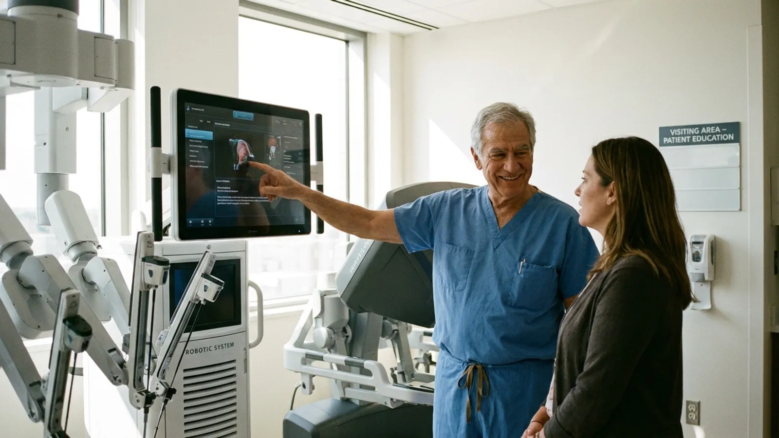
Retinal Detachment Surgery: Symptoms, Repair Techniques, Recovery, and Costs
Recovery after retinal detachment surgery requires strict head positioning, travel restrictions with gas bubbles, and careful monitoring during healing.

Retinal Detachment Surgery: Symptoms, Repair Techniques, Recovery, and Costs
Quick Answer: Retinal detachment surgery is an urgent ophthalmic intervention that reattaches the neurosensory retina to the underlying tissue to prevent severe, irreversible visual loss. Surgery is needed as soon as possible, ideally within 24 hours, while the central macula is still attached; once the macula has detached, it is still done within a few days, without delay. Treatment success depends primarily on prompt clinical presentation, whether the macula remains attached, appropriate surgical selection (such as vitrectomy or scleral buckle), and strict adherence to postoperative head positioning.
Key Takeaways:
- Sudden flashes of light, a rapid increase in dark floaters, or a peripheral curtain-like shadow represent an ocular emergency requiring same-day dilated examination.
- Preserving baseline visual acuity depends heavily on repairing the retina before the central macula detaches (macula-on status).
- Modern repair techniques include pars plana vitrectomy, scleral buckling, and pneumatic retinopexy, often supported by laser photocoagulation or cryopexy.
- Postoperative head positioning is required when gas or silicone oil tamponades are placed in the eye; the surgeon decides the position based on where the tear is, and with a superior tear, sitting upright may be enough.
- Air travel and ascent to high altitudes are strictly prohibited while an intraocular gas bubble remains in the eye.
A retinal detachment occurs when the neurosensory retina separates from the underlying retinal pigment epithelium at the back of the eye, depriving photoreceptors of oxygen and essential nutrients. Left untreated, progressive detachment causes cellular damage within the light-sensitive tissue, leading to profound, irreversible vision loss in the affected eye.
If you notice sudden flashes, a shower of new floaters or a shadow in your vision, get an eye examination the same day where you are and do not delay treatment in order to travel; going abroad makes more sense for planned, non-urgent care or a second opinion.
What Is Retinal Detachment and Who Faces the Highest Risk?
Retinal detachment occurs when the light-sensitive neurosensory retina separates from the underlying retinal pigment epithelium at the back of the eye. Individuals with severe myopia (high nearsightedness), prior cataract surgery, ocular trauma, lattice degeneration, or a personal or family history of retinal breaks face the greatest clinical risk.
The eye functions much like an optical camera: the clear cornea and crystalline lens focus incoming light onto the retina lining the rear inner wall. The retina converts light rays into neural signals sent through the optic nerve to the visual cortex of the brain. When physical separation occurs, subretinal fluid accumulates in the space beneath the sensory layer.
Clinicians classify retinal detachment into three primary mechanisms:
- Rhegmatogenous Detachment: The most common form, arising from a full-thickness break, tear, or hole in the retina. Liquefied vitreous gel passes through the opening into the subretinal space, peeling the sensory retina away from the underlying vascular bed.
- Tractional Detachment: Occurs when contractile fibrous membranes on the retinal surface pull the tissue forward. This is most frequently seen in advanced proliferative diabetic retinopathy, retinal vein occlusions, or retinopathy of prematurity.
- Exudative (Serous) Detachment: Involves fluid pooling beneath an intact retina due to inflammatory disorders, hydrostatic imbalances, choroidal tumors, or severe systemic hypertension.
Several anatomical and medical factors elevate an individual's baseline risk of developing a rhegmatogenous retinal break:
- High Myopia: Eyes with axial lengths exceeding 26 mm have thinned, stretched retinal tissue that is structurally vulnerable to peripheral tears.
- Prior Intraocular Surgery: Procedures such as cataract extraction alter vitreous dynamics, moderately increasing the lifetime risk of posterior vitreous detachment (PVD) and subsequent retinal tears.
- Peripheral Retinal Degenerations: Conditions like lattice degeneration cause localized retinal thinning and strong vitreoretinal adhesions.
- Direct Ocular Trauma: Blunt or penetrating facial injuries can trigger immediate or delayed tears through mechanical shock.
- Fellow Eye History: A patient who has had a retinal detachment in one eye carries a reported 10% to 15% lifetime probability of developing a tear or detachment in the other eye.
Retinal Detachment Symptoms and Treatment Urgency: When to Seek Emergency Care
A sudden onset of light flashes (photopsia), a dramatic increase in dark floaters, or a dark shadow spreading across the visual field signals an ocular emergency requiring same-day evaluation. When the central retina (macula) remains attached (macula-on), surgical intervention as soon as possible, ideally within 24 hours, offers the highest likelihood of preserving fine central vision.
The earliest phase of retinal tear formation often produces mechanical traction on the neurosensory layer. Because the retina lacks pain receptors, these events occur without physical discomfort, making visual symptoms the sole warning signs. Recognizing the typical retinal detachment symptoms and treatment urgency can mean the difference between preserving functional vision and experiencing significant visual impairment.
Visual Symptom Progression
- Microscopic Vitreous Separation: Photopsia (light flashes) and subtle micro-floaters appear as the vitreous gel pulls against the retina.
- Full-Thickness Retinal Tear: A sudden shower of dark floaters ("soot" or "cobwebs") develops, often caused by microscopic bleeding from a torn retinal vessel.
- Progressive Subretinal Fluid Accumulation: A dark curtain or peripheral shadow moves inward from one side of the visual field.
- Macular Involvement: Distorted, blurred, or lost central vision occurs as fluid undermines the central retina.
Key symptoms include:
- Photopsia (Light Flashes): Streaks of light, lightning-like arcs, or brief sparks visible in peripheral vision, most noticeable in dim environments or during rapid eye movements.
- Floater Showers: Sudden bursts of dozens or hundreds of black spots, thread-like strands, or cobwebs.
- Peripheral Visual Field Defect: A progressive gray or dark curtain moving inward from any direction (top, bottom, or sides), representing detached peripheral retina.
- Distorted Central Acuity (Metamorphopsia): Wavy lines, visual graying, or sudden loss of sharpness indicating that fluid has advanced under the macula.
The clinical urgency of surgical repair depends heavily on macular status:
- Macula-On Detachment: The patient retains crisp central vision (often 20/20 to 20/40), but the detached peripheral tissue threatens to spread across the center. Surgery is performed as soon as possible, ideally within 24 hours, to secure the tissue before central vision is compromised.
- Macula-Off Detachment: The macula has detached, causing significant loss of central vision. Surgery is still urgent and is usually performed within a few days, without delay; because the central photoreceptors have already separated, visual recovery occurs gradually over several months.
Who Should You Consult: General Ophthalmologist or Vitreoretinal Specialist?
While an emergency physician, optometrist, or general ophthalmologist can perform an initial dilated eye examination, surgical repair requires a fellowship-trained vitreoretinal specialist. Patients presenting with acute detachment symptoms should seek immediate referral to a dedicated retina clinic or an eye emergency center equipped for vitreoretinal surgery.
An optometrist or general ophthalmologist uses a slit lamp and indirect ophthalmoscopy with scleral depression to examine the entire retinal periphery. If a detachment, tear, or unexplained dense vitreous hemorrhage is identified, they refer the patient directly to a vitreoretinal surgeon.
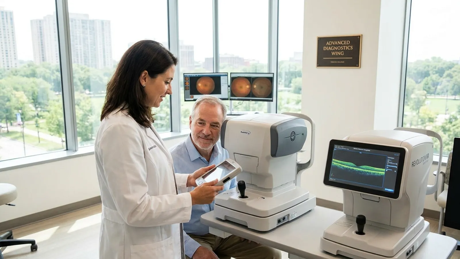
Vitreoretinal specialists complete advanced multi-year surgical fellowships focusing on micro-incisional surgery within the posterior segment of the eye. They rely on specialized diagnostic equipment to map the detachment:
- Widefield Fundus Photography: High-resolution digital imaging that captures up to 200 degrees of the retina in a single view to document breaks.
- Optical Coherence Tomography (OCT): Cross-sectional, micron-level scans that confirm whether subretinal fluid has elevated the foveal center of the macula.
- B-Scan Ocular Ultrasound: High-frequency acoustic imaging used when dense vitreous hemorrhage or mature cataracts prevent clear optical views of the fundus.
What Is the Treatment for a Detached Retina? Surgical Options and Contraindications
Primary surgical interventions include pars plana vitrectomy, scleral buckling, and pneumatic retinopexy, often combined with laser photocoagulation or cryopexy. Contraindications depend on anatomical factors: pneumatic retinopexy is unsuitable for inferior tears or severe proliferative vitreoretinopathy (PVR), while patients unable to maintain prone positioning may require alternative tamponade choices.
When evaluating what is the treatment for a detached retina, the ophthalmic surgeon selects a technique based on tear location, ocular anatomy, lens status (phakic versus pseudophakic), and the presence of scar tissue.
| Surgical Method | Primary Indications | Invasiveness & Setting | Tamponade / Fixation Used | Common Contraindications |
|---|---|---|---|---|
| Pneumatic Retinopexy | A single break, or a group of breaks within one clock hour, in the upper two-thirds of the retina (between the 8 and 4 o'clock positions) | Outpatient clinic procedure | Intraocular expandable gas bubble (SF6 or C3F8) with cryopexy or laser | Inferior tears (4:00 to 8:00), severe PVR, significant media opacities |
| Scleral Buckling | Young phakic patients, inferior breaks, dialyses, round holes without severe PVR | Operating room (outpatient or short stay) | External silicone sponge or solid band sutured to sclera | Extensive posterior breaks, severe scleral thinning, previous buckle complications |
| Pars Plana Vitrectomy (PPV) | Complex detachments, posterior breaks, vitreous hemorrhage, pseudophakic eyes | Operating room micro-incisional surgery (23G, 25G, or 27G) | Internal gas (SF6/C3F8) or medical-grade silicone oil | Inability to comply with any head positioning (relative; silicone oil may adapt) |
| Combined Buckle-Vitrectomy | Severe PVR, recurrent detachments, giant retinal tears, high-trauma cases | Comprehensive operating room procedure | External silicone band plus internal gas or silicone oil | Extreme surgical fragility, unmanageable systemic medical instability |
Pars Plana Vitrectomy (PPV)
Vitrectomy surgery is the most common intervention for retinal reattachment in modern ophthalmic practice. The surgeon creates three microscopic self-sealing incisions through the pars plana, the safe entry zone in the white sclera:
- One port infuses balanced salt solution to maintain stable intraocular pressure.
- The second port holds an endoilluminator fiber-optic light.
- The third port accommodates high-speed vitrector probes (cutting up to 10,000 to 20,000 cuts per minute), micro-forceps, or endolaser probes.
The surgeon cuts and removes the vitreous gel, relieves all vitreoretinal traction, flattens the retina using heavy liquid or air-fluid exchange, applies laser photocoagulation around all breaks, and fills the vitreous cavity with an expandable gas mixture or silicone oil. For more detail on this operation and its recovery, see vitrectomy for retinal detachment.
Scleral Buckle
A scleral buckle involves securing a flexible silicone band, tire, or sponge around the exterior wall of the eye under the conjunctiva and extraocular muscles. By indenting the sclera inward, the buckle relieves mechanical vitreous pull and brings the pigment epithelium into direct contact with the retinal tear. Cryopexy (freezing therapy) applied before placing the band stimulates a localized adhesion that seals the defect.
Pneumatic Retinopexy
Pneumatic retinopexy is a minimally invasive, office-based procedure. The surgeon injects a small bubble of expandable gas (such as sulfur hexafluoride [SF6] or perfluoropropane [C3F8]) directly into the vitreous cavity. The patient positions their head so the floating bubble presses directly against the retinal tear, closing it and allowing the retinal pigment epithelium to resorb the subretinal fluid over 24 to 48 hours. Cryopexy is performed before the gas injection, or laser photocoagulation is applied once the retina flattens.
Retinal Adhesion and Tamponade Agents
Mechanical reattachment alone is insufficient; stable chorioretinal adhesions must form around the tear margins:
- Laser Photocoagulation: Uses focused thermal energy to create microscopic burns that heal into firm pigmented scars within 10 to 14 days.
- Cryopexy: Uses a freezing probe on the outer eye wall to generate a cold-induced scar around peripheral breaks.
- Gas Tamponades (SF6, C3F8): Temporary agents that dissolve on their own, SF6 in about 2 to 3 weeks and C3F8 in about 6 to 8 weeks, requiring no secondary removal surgery.
- Silicone Oil Tamponade: Used for complex detachments, giant tears, or patients unable to maintain positioning. Silicone oil does not dissolve on its own and requires a secondary surgical procedure for removal once the retina is stable (typically 3 to 6 months later).
What to Expect During the Procedure: Anesthesia and Essential Questions for Your Surgeon
Retinal detachment surgery is typically performed as an outpatient procedure under local anesthesia with intravenous sedation or under general anesthesia, depending on surgical complexity and patient health. Before surgery, patients should discuss macula status, the choice of tamponade agent (gas or silicone oil), expected recovery milestones, and potential risks with their surgeon.
Most adults undergoing vitrectomy or scleral buckling receive local anesthesia with intravenous sedation. An ophthalmic anesthesiologist administers sedatives to keep the patient calm and comfortable, followed by a retrobulbar or peribulbar block: a local anesthetic injection behind or around the eye that numbs the globe and temporarily stops eye movement. Under this block, patients do not feel sharp pain during the operation.
General anesthesia is typically selected for extensive combined procedures, pediatric patients, individuals with spinal conditions preventing supine positioning, or patients with severe claustrophobia or anxiety.
Before entering the operating room, patients can clarify key clinical parameters by asking their surgeon:
- Is my central macula fully attached, partially detached, or completely off?
- Which surgical technique is best suited for my retinal tear configuration, and why?
- Will you be using an intraocular gas bubble or silicone oil as an internal tamponade?
- What specific head positioning will be required, and for how many hours per day?
- What is the anticipated visual recovery timeline based on my eye's presentation?
- What specific symptoms should prompt an immediate after-hours call?
Recovery After Retinal Detachment Surgery: Positioning and Healing Timelines
Postoperative recovery centers on maintaining the head position your surgeon prescribes for several days to weeks when an intraocular gas bubble or silicone oil is placed to support the retina. Initial surface healing and bubble reabsorption occur over several weeks, while visual stabilization typically continues over 3 to 6 months.
Adherence to head positioning (posturing) directly influences the primary success rate of vitrectomy surgery. Because gas and oil are lighter than fluid, they float upward within the eye. The patient must position their head so the buoyant surface of the bubble presses precisely against the healing retinal breaks. Your surgeon decides the exact position based on where the tear is; with a superior tear, sitting upright may be enough.
Critical Postoperative Care Phases
Days 1 to 7 (Acute Positioning and Protection):
- Patients keep the position their surgeon prescribes (for example facedown, side-lying, or chin-down) for up to 16 to 20 hours per day. Specialized support equipment (cushioned chairs, face cradles, prism glasses) helps reduce neck and shoulder strain.
- A rigid eye shield is taped over the operative eye during sleep to prevent accidental rubbing or pressure.
- Topical drop regimens begin: broad-spectrum antibiotics for 7 to 10 days, corticosteroids for 4 to 6 weeks to control intraocular inflammation, and cycloplegics to manage ciliary muscle spasm.
Weeks 2 to 4 (Intermediate Healing):
- The gas bubble gradually shrinks. Patients notice a horizontal line across their vision that drops like a water level, with clearer vision appearing above the line.
- Light daily tasks resume. Heavy physical exertion, bending below the waist, and lifting objects heavier than 5 kg (11 lbs) remain restricted.
Months 2 to 6 (Visual Stabilization and Refraction):
- Any remaining gas bubble has fully resorbed by now (SF6 usually within 2 to 3 weeks, C3F8 within 6 to 8 weeks), replaced naturally by aqueous humor.
- If silicone oil was placed, a planned outpatient procedure is scheduled to remove the oil once the retina is secure.
- Visual acuity settles gradually. A final prescription for eyeglasses is determined once corneal shape and macular thickness stabilize.
What Are the Long Term Restrictions After Retinal Detachment Surgery?
Patients with an intraocular gas bubble are strictly prohibited from flying, traveling to high altitudes, or undergoing nitrous oxide anesthesia due to the risk of severe intraocular pressure spikes. Once fully healed, most patients resume daily activities, though high-impact contact sports and activities carrying elevated eye trauma risks require protective eyewear.
Understanding what are the long term restrictions after retinal detachment surgery helps prevent sight-threatening complications both during the initial gas phase and after structural reattachment is complete.
Absolute Immediate Contraindications (While Gas Bubble Remains)
- Aviation / Flying: Atmospheric cabin pressure in commercial aircraft drops at cruising altitude. Under reduced ambient pressure, an intraocular gas bubble expands rapidly, which can cause acute ocular hypertension, central retinal artery occlusion, and severe, irreversible vision loss within hours.
- High-Altitude Travel: Driving through mountain passes or visiting locations significantly above sea level presents the same gas-expansion risks as flying.
- Nitrous Oxide Anesthesia: Nitrous oxide diffuses rapidly into gas-filled spaces, causing dangerous bubble expansion and intraocular pressure spikes. Patients should wear a medical alert wristband stating that an intraocular gas bubble is present.
- Sleeping and Resting Position: Do not lie on your back unless your surgeon tells you to.
- Driving: Do not drive until your vision allows it and your surgeon has cleared you.
Long-Term Activity and Eye Health Guidelines
Once the retina is stable and the gas bubble has resolved, patients can safely return to most routine physical activities:
- Contact and High-Impact Sports: Martial arts, boxing, rugby, and racquet sports carry risks of direct ocular impact. Polycarbonate protective sports goggles (meeting ASTM F803 standards) should be worn during racquet sports, basketball, and workshop activities.
- High-G-Force Activities: Bungee jumping, skydiving, and intense roller coasters involve rapid acceleration forces that place stress on vitreoretinal interfaces and should be approached with caution.
- Routine Exercise: Running, swimming, gym workouts, and low-impact fitness activities can typically resume 4 to 6 weeks after surgery, following the surgeon's clearance.
- Monitoring the Fellow Eye: Because the risk of retinal breaks in the opposite eye is elevated, prompt reporting of new floaters or flashes remains essential.
Retinal Detachment Surgery Cost and International Patient Considerations
The cost of retinal detachment surgery varies widely based on geographic region, hospital facility fees, surgeon expertise, and the required technique. In private international healthcare settings, comprehensive vitreoretinal surgery packages often range from $2,500 to $6,000 USD (£2,000 to £4,800 GBP / €2,300 to €5,500 EUR), compared to significantly higher out-of-pocket costs in the United States.
A new retinal detachment should be treated without delay where you are, rather than postponed to arrange travel; treatment abroad is better suited to planned, non-urgent care, such as a planned second operation or a second opinion from a specialist ophthalmology team.
Managing the logistical and financial aspects of planned ophthalmic care requires a clear breakdown of costs and careful travel planning:
| Healthcare System / Country | Estimated Out-of-Pocket / Private Cost Range (USD) | Estimated Cost Range (GBP / EUR) | Key Logistical and Clinical Factors |
|---|---|---|---|
| United States (Self-Pay / Uninsured) | $8,000-$18,000+ | £6,400-£14,400 / €7,400-€16,500 | Variable facility fees; separate surgeon and anesthesia billing. |
| United Kingdom (Private Track) | $4,500-$8,500 | £3,500-£6,500 / €4,100-€7,800 | Fixed-price consultant packages; emergency care is available via the NHS. |
| European Medical Hubs (e.g., Spain, Germany) | $4,000-$7,500 | £3,200-£6,000 / €3,700-€7,000 | Advanced vitreoretinal units; pricing typically bundled into a single hospital fee. |
| International Hubs (e.g., Turkey, Czechia) | $2,500-$5,500 | £2,000-£4,400 / €2,300-€5,100 | Package contents vary by hospital. |
These are indicative ranges that vary by clinic and by case; they usually do not include accommodation or the cost of a possible second operation.
For international patients traveling for private eye surgery, safety precautions regarding travel are paramount:
- Travel Restrictions After Gas Injection: Patients who receive intraocular gas (SF6 or C3F8) cannot fly until the gas bubble has completely resorbed. This means staying near the hospital, without traveling to higher altitudes, for about 2 to 3 weeks with SF6 or 6 to 8 weeks with C3F8, until an eye examination confirms the gas has gone.
- Silicone Oil and Flying: For complex or recurrent detachments, your surgeon may choose silicone oil instead of gas. Silicone oil does not expand with atmospheric pressure changes, so flying is possible, but it is a medical choice rather than a travel option: it needs a second operation to remove it, usually 3 to 6 months later, and it can raise eye pressure and cause cataract.
- Package Inclusions: Comprehensive retinal detachment surgery cost quotes should itemize preoperative OCT and ultrasound diagnostics, operating room fees, surgeon and anesthesia charges, tamponade supplies, postoperative protective shields, and follow-up examinations within the first month.
- Follow-Up at Home: Before you fly home, arrange care with a local ophthalmologist and take your operation report with you. Plan regular check-ups during the first weeks after you return, because proliferative vitreoretinopathy (PVR), the scarring that can detach the retina again, usually appears within 6 to 12 weeks of surgery.
Risks and Potential Complications of Vitreoretinal Surgery
Complications following retinal reattachment surgery include redetachment, elevated intraocular pressure, cataract progression, endophthalmitis, and proliferative vitreoretinopathy (PVR). Most surgical complications are manageable with timely intervention, though some may require secondary procedures to stabilize visual acuity.
While modern vitreoretinal surgery achieves anatomical reattachment in reported ranges of 85% to 90% of primary interventions, patients should understand potential risks:
- Proliferative Vitreoretinopathy (PVR): The leading cause of surgical redetachment, occurring in approximately 5% to 10% of cases. Cellular membranes grow on the retinal surface, contracting and pulling the retina away from the eye wall, which requires repeat surgery.
- Secondary Retinal Redetachment: May develop due to missed peripheral tears, newly formed breaks, or persistent vitreoretinal traction, requiring repeat vitrectomy or scleral buckle revision.
- Accelerated Cataract Progression: In patients with their natural lens intact (phakic eyes), vitrectomy accelerates nuclear sclerotic cataract formation. Most phakic patients over age 50 require routine cataract surgery within 6 to 18 months following vitrectomy.
- Ocular Hypertension and Glaucoma: Postoperative inflammation, gas bubble expansion, or steroid eye drop use can elevate intraocular pressure, which is typically managed with topical pressure-lowering drops.
- Endophthalmitis: A rare (<0.1%) but serious intraocular infection that requires emergency intravitreal antibiotic injections.
- Corneal Surface Irregularities: Temporary surface abrasions or corneal haze that typically resolve with lubricating ointments and temporary bandage contact lenses.
Urgent warning signs after surgery: contact your surgeon or an eye emergency service straight away if you develop severe or worsening eye pain, nausea or vomiting, worsening vision, a new curtain or shadow in your vision, or increasing redness or discharge.
Frequently Asked Questions
Are you awake for retinal detachment surgery?
Yes, most adult patients are awake but deeply relaxed under local anesthesia with monitored intravenous sedation. A local nerve block completely numbs the eye and temporarily stops eye movement, ensuring you do not feel sharp pain during the operation. General anesthesia is typically reserved for pediatric patients, long or complex surgical cases, or patients with severe anxiety or medical conditions that prevent them from lying flat.
How long after retinal detachment surgery can I fly?
You cannot fly as long as an intraocular gas bubble remains in your eye, which typically takes about 2 to 3 weeks for SF6 gas and 6 to 8 weeks for C3F8 gas. The drop in cabin pressure at flight altitudes causes the gas bubble to expand rapidly, which can lead to dangerously high intraocular pressure and severe vision loss. If your surgeon uses silicone oil instead of a gas bubble, you can safely fly once cleared during your early postoperative checkups.
How is treatment for detached retina in the UK managed between the NHS and private providers?
When seeking treatment for detached retina, UK pathways include both public and private options. Under the NHS, emergency care is delivered through hospital eye casualty units with zero out-of-pocket cost, triaging patients based on clinical urgency (macula-on versus macula-off). Private ophthalmic care (£3,500 to £6,500) provides direct access to a chosen vitreoretinal consultant, flexible operating theater scheduling, and private clinic amenities while following standard clinical safety protocols.
Can a retinal detachment heal on its own without surgery?
No, a full neurosensory retinal detachment cannot heal on its own. Because subretinal fluid continually enters through the retinal tear, the detachment progressively enlarges until it causes total loss of functional vision in that eye. A retinal tear found before the retina detaches is usually treated in the clinic with laser or freezing treatment (cryopexy); this is the standard treatment and greatly lowers the chance that the tear progresses to a detachment. Once fluid has collected under the retina, it requires prompt surgical repair to reattach the retina to its underlying vascular supply.
Not sure which hospital is right for your case?
Upload your medical reports and our AI will match them against the verified capabilities of every hospital in our network.
- Upload your reports (no account needed)
- Our AI matches your case with hospitals that can actually treat it
- You choose who receives your case. We present it in the hospital’s own language, and the hospital replies directly to you
Free and anonymous. Your documents stay private until you decide to send them.