Robotic Surgery for Rectal Cancer: Is a Permanent Colostomy Inevitable?
Robotic surgery for rectal cancer aims to preserve sphincter function and may help avoid a permanent colostomy in suitable candidates using 3D visualization.

Robotic Surgery for Rectal Cancer: Is a Permanent Colostomy Inevitable?
Quick Answer: A permanent colostomy is not inevitable for most patients with rectal cancer. Advanced robotic surgery, precise total mesorectal excision (TME), and pre-operative therapies allow surgeons to spare the anal sphincter and preserve natural bowel continuity in the vast majority of mid-to-low rectal cases.
Key Takeaways:
- Advanced robotic platforms provide three-dimensional visualization and articulated instruments that allow precise dissection within the deep pelvis, helping surgeons spare pelvic nerves and natural sphincter muscles.
- Sparing the sphincter requires an adequate distal margin below the tumor without direct invasion into the external sphincter muscle or pelvic floor.
- A temporary loop ileostomy is often used for 8 to 12 weeks to protect low pelvic joins during healing, which is entirely distinct from a permanent colostomy.
- Low Anterior Resection Syndrome (LARS) affects bowel frequency and urgency post-surgery, though symptoms typically settle over time with pelvic rehabilitation, medication, and dietary adjustments.
- International medical centres in Turkey offer robotic rectal cancer resections in Joint Commission International (JCI)-accredited facilities at roughly 60% to 75% lower cost than private care in the US or UK.
Robotic surgery for rectal cancer involves using computerized, surgeon-controlled mechanical instruments to remove malignant tumors from the rectum-the lower portion of the large intestine-with high anatomical precision. By combining miniature wristed instruments with high-magnification optical systems, this minimally invasive approach allows colorectal specialists to navigate the rigid confines of the human pelvis, removing malignant tissue along natural anatomical planes while safeguarding the nerves and muscles responsible for urinary, sexual, and fecal continence.
Is a Permanent Colostomy Inevitable with Rectal Cancer?
A permanent colostomy is not inevitable for most rectal cancer patients; modern robotic techniques, accurate tumor staging, and pre-operative chemoradiation allow the majority of mid-to-low rectal tumors to be removed while preserving the anal sphincter and natural bowel continuity.
Historically, rectal cancers situated in the lower third of the rectum (within 5 centimeters of the anal verge) almost always required an abdominoperineal resection (APR). This operation removes the entire rectum, anal canal, and sphincter complex, leaving the patient with a lifelong end-colostomy. Today, high-resolution magnetic resonance imaging (MRI) staging, total neoadjuvant therapy (TNT) to downsize tumors before surgery, and refined surgical instrumentation allow surgeons to achieve rectal cancer surgery without permanent bag placement in many patients who previously faced total sphincter removal.
Preserving the sphincter requires three core oncological criteria:
- The cancer must not directly invade the external anal sphincter or the puborectalis/levator ani muscle complex.
- The surgeon must be able to achieve a microscopically clear distal margin (the healthy tissue below the tumor).
- The patient must have adequate baseline sphincter function to ensure post-surgical continence is functional.
When these conditions are met, surgeons perform a low anterior resection (LAR) or ultra-low anterior resection with a coloanal anastomosis. In these procedures, the diseased section of the bowel is excised, and the remaining healthy colon is joined directly to the lower rectum or anal canal, preserving the natural route for waste elimination.
How Robotic Technology Protects Sphincter Function in Low Pelvic Tumors
Robotic platforms provide 3D high-definition visualization and articulated instruments with seven degrees of freedom, allowing surgeons to dissect within the narrow bony pelvis with sub-millimeter precision, sparing autonomic nerves responsible for urinary, sexual, and continence function.
The human pelvis is a tight, conical space bounded by rigid bone. In conventional open surgery, access to the deep pelvis requires large incisions and forceful retraction, which can compromise visibility. Standard laparoscopy provides minimally invasive access, but straight, rigid instruments lack the dexterity needed to work around the tight curve of the sacrum.
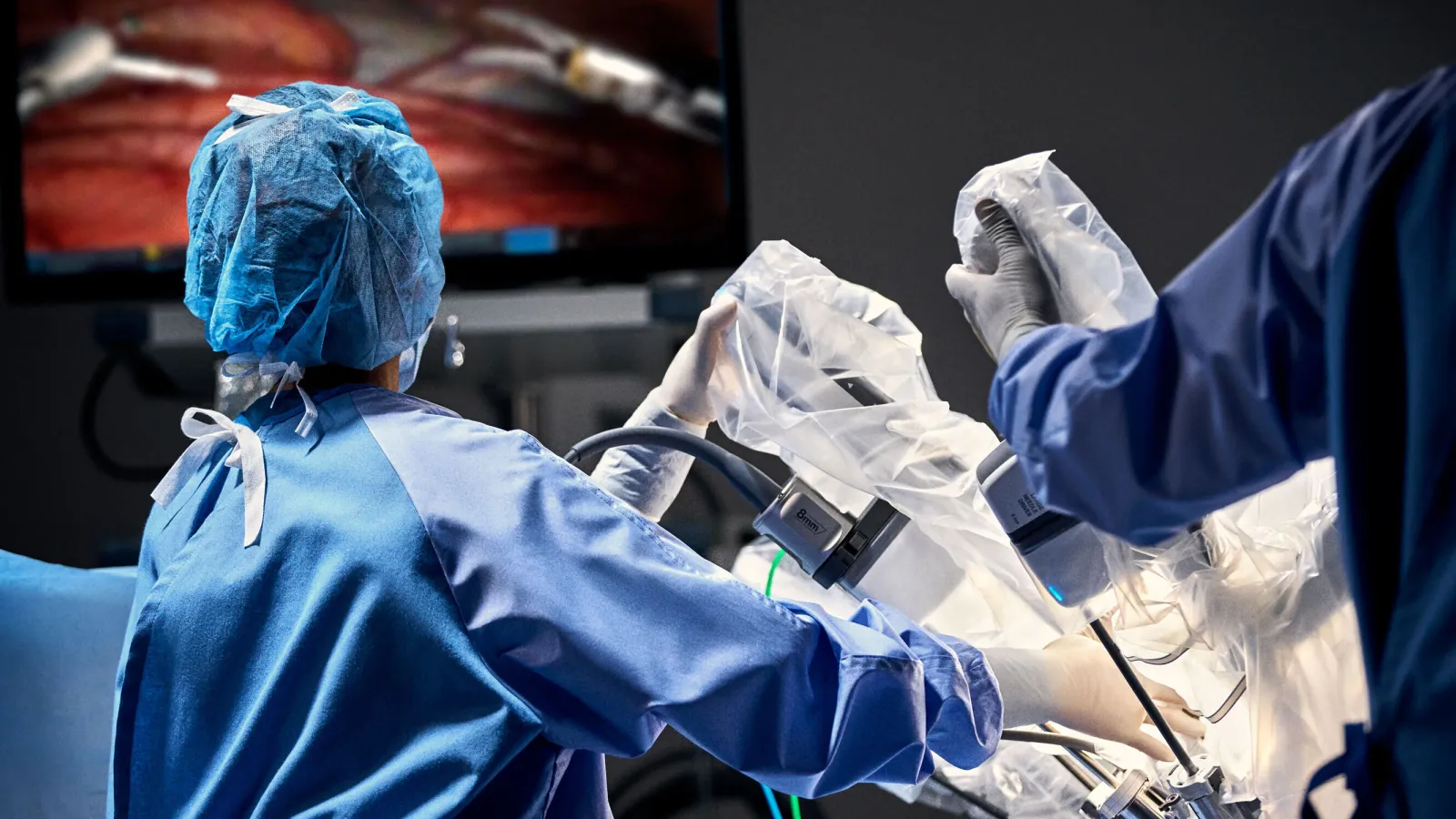
Systems such as the da Vinci surgical platform, cleared by the US FDA in the United States for general and colorectal procedures, address these anatomical challenges through specific technical advantages:
- Tremor Filtration and Scaling: Micro-movements of the surgeon's hands are translated into steady, scaled mechanical actions, preventing accidental tissue trauma.
- EndoWrist Articulation: Instruments articulate beyond the range of the human wrist, allowing circumferential dissection around low tumors.
- Stable 3D High-Definition Optics: The camera system delivers magnified, stereoscopic views of tissue planes, distinguishing between cancerous boundaries, blood vessels, and fine autonomic nerve plexuses.
- Ergonomic Access: Surgeons can reach the pelvic floor muscles without exerting torque on surrounding healthy tissues.
By improving visibility and reach in the narrow pelvis, da Vinci robotic surgery rectal cancer procedures help surgeons preserve the pelvic autonomic nerves (the hypogastric and pelvic splanchnic nerves). Sparing these nerve networks reduces the risk of long-term bladder dysfunction, sexual dysfunction, and internal sphincter dysmotility.
Anatomical Criteria: Tumor Height, Margins, and Total Mesorectal Excision (TME)
Sphincter preservation depends primarily on achieving a clear distal margin-typically 1 to 2 centimeters below the tumor edge-and maintaining an intact mesorectal envelope through total mesorectal excision (TME) without oncological compromise.
The feasibility of sphincter-preserving surgery rectal cancer protocols is governed by tumor location and its relationship to the mesorectum-the fatty tissue sheath surrounding the rectum that houses blood vessels and lymph nodes. Total Mesorectal Excision (TME) is the standard surgical technique for rectal cancer; it requires removing this entire fatty envelope with intact, smooth fascial margins to minimize local recurrence.
| Rectal Zone | Distance from Anal Verge | Standard Surgical Approach |
|---|---|---|
| Upper Rectum | 10-15 cm | Standard Anterior Resection |
| Mid Rectum | 5-10 cm | Low Anterior Resection (TME) |
| Low Rectum | < 5 cm | Ultra-Low LAR / Intersphincteric Resection |
| Anal Canal | 0-2 cm | Intersphincteric Resection or APR |
Key anatomical factors evaluated during pre-operative pelvic MRI include:
- Tumor Height: Measured from the anal verge. High (10-15 cm) and mid-rectal (5-10 cm) tumors almost always allow sphincter preservation. Low rectal tumors (<5 cm) require specialized ultra-low or intersphincteric techniques.
- Distal Resection Margin (DRM): While historical guidelines required a 5 cm margin, contemporary clinical data demonstrates that a 1 to 2 cm margin-and in select ultra-low cases after radiation response, a sub-centimeter margin-is oncologically safe without increasing recurrence rates.
- Circumferential Resection Margin (CRM): The distance between the deepest tumor invasion and the outer boundary of the mesorectal fascia. A clear CRM (>1 mm) is vital for local cancer control.
- Intersphincteric Plane: For tumors touching the internal sphincter, surgeons can dissect between the internal and external sphincter muscles (intersphincteric resection), removing the internal muscle while preserving the outer striated muscle that maintains voluntary continence.
Temporary Diverting Ileostomy vs. Permanent Colostomy: What to Expect
A temporary protective ileostomy diverts stool for 8 to 12 weeks while the low pelvic anastomosis heals securely, after which it is surgically reversed, distinct from a permanent end-colostomy where the rectum and sphincter complex are entirely excised.
Patients preparing for rectal cancer surgery without colostomy are often surprised to learn they may still wake up with a stoma bag. It is essential to distinguish between a temporary loop ileostomy and a permanent end-colostomy.
When a low or ultra-low anastomosis is constructed close to the anus, the newly joined tissues are vulnerable to pressure and bacterial contamination from passing stool. A temporary loop ileostomy brings a small loop of the small intestine (ileum) through the abdominal wall to redirect digestive waste into an external pouch. This protects the pelvic join, significantly reducing the clinical consequences of an anastomotic leak.
| Feature | Temporary Diverting Ileostomy | Permanent End-Colostomy |
|---|---|---|
| Intestinal Source | Small intestine (Ileum) | Large intestine (Colon) |
| Location on Abdomen | Usually lower right quadrant | Usually lower left quadrant |
| Stool Consistency | Liquid to paste-like | Formed to semi-formed |
| Duration | 8 to 12 weeks (up to 6 months if post-op chemo is needed) | Lifelong |
| Sphincter Status | Anal sphincter remains intact and functional | Sphincter complex is entirely excised |
| Second Surgery | Minor reversal surgery to close the stoma | None (stoma is permanent) |
| Primary Indication | Protects low pelvic connection (anastomosis) | Performed when tumor invades sphincter muscles |
Once clinical evaluation or imaging (such as a water-soluble contrast enema) confirms that the pelvic connection has healed without leakage, a brief, minor surgical procedure is performed to reconnect the small bowel and close the abdominal wall opening.
Recovery Timeline and Managing Bowel Function After Surgery
Hospital recovery after robotic rectal resection typically spans 4 to 7 days, with complete surgical recovery taking 6 to 8 weeks, during which dietary adjustments and pelvic floor physical therapy help manage temporary changes in bowel frequency known as Low Anterior Resection Syndrome (LARS).
During recovery after rectal cancer surgery, the body requires time to heal both the surgical incisions and the modified digestive tract. Removing part or all of the rectal reservoir alters how the body stores and evacuates stool.
Milestones of Recovery
- Days 1-2: Early ambulation begins; clear liquids progress to a low-residue, soft diet; pain is managed primarily through non-opioid multimodal analgesia.
- Days 3-5: Stoma education and self-management training occur; urinary catheters are removed; bowel function (stoma output or natural passage) is monitored.
- Weeks 2-6: Baseline energy returns; light daily activity resumes; heavy lifting (>10 lbs / 4.5 kg) is avoided to protect abdominal incisions.
- Weeks 8-12: Follow-up imaging evaluates the internal anastomosis; planning for temporary stoma reversal occurs if systemic chemotherapy is completed or not required.
Understanding and Managing LARS
When bowel continuity is restored, many patients experience Low Anterior Resection Syndrome. Symptoms can include increased bowel frequency, urgency, clustering (multiple small bowel movements over a few hours), and difficulty distinguishing gas from stool.
LARS symptoms are most pronounced in the first 3 to 6 months and typically improve steadily over 1 to 2 years. Management strategies include:
- Dietary Titration: Consuming small, frequent meals low in insoluble fiber, caffeine, and simple sugars while using soluble fiber supplements to add bulk.
- Medications: Using anti-motility agents (such as loperamide) under medical supervision to slow transit time and improve stool consistency.
- Pelvic Floor Physiotherapy: Targeted biofeedback and pelvic floor exercises to strengthen the external sphincter and improve voluntary control.
Robotic Rectal Cancer Surgery Costs: Turkey vs. US and UK
Robotic rectal cancer surgery in Turkey typically ranges from $9,000 to $16,000 (£7,200 to £12,800), including pre-operative imaging, robotic operative fees, and multi-day inpatient stays, compared to $45,000-$85,000 in the United States and £22,000-£35,000 privately in the United Kingdom.
The total rectal cancer surgery cost varies widely depending on the country of treatment, hospital infrastructure, surgical technology fees, and the complexity of postoperative care. Robotic procedures carry higher equipment and consumable costs (robotic arms, specialized staplers, visual cartridges) than conventional open surgery, which often drives up private pricing in Western systems.
| Country | Estimated Cost Range (USD) | Estimated Cost Range (GBP) | Typical Inclusions |
|---|---|---|---|
| United States (Self-Pay) | $45,000 - $85,000+ | £36,000 - £68,000+ | Surgeon, robotic facility fee, 4-6 day stay (tests billed separately) |
| United Kingdom (Private) | $28,000 - $44,000 | £22,000 - £35,000 | Hospital package, surgeon/anesthetist fees, initial recovery |
| Turkey (Accredited Centers) | $9,000 - $16,000 | £7,200 - £12,800 | Comprehensive package: pre-op MRI/CT, robotic TME, pathology, 5-7 hospital days, transfers |
The competitive robotic rectal cancer surgery cost in Turkey is driven by lower facility operating overhead and favorable exchange rates rather than differences in medical technology. Leading Turkish comprehensive cancer centers utilize modern da Vinci surgical systems and maintain international accreditations, such as Joint Commission International (JCI) accreditation or ISO certifications. For international patients, these packages frequently include airport transfers, multilingual patient coordinators, and dedicated postoperative nursing care.
Patient Candidacy: Are You Eligible for Robotic Sphincter Preservation?
Eligibility for sphincter-sparing surgery requires a tumor that does not invade the external anal sphincter or levator ani muscles, acceptable baseline continence, and favorable response to neoadjuvant therapy when indicated.
Determining whether a patient is a suitable candidate for low rectal cancer surgery with sphincter preservation involves a multidisciplinary tumor board review consisting of colorectal surgeons, medical oncologists, radiation oncologists, and gastrointestinal radiologists.
Ideal Candidates for Sphincter Preservation
- Patients with tumors in the upper, middle, or lower rectum where a distal margin of at least 1 cm (or clear margin confirmed via frozen section) is achievable above the sphincter complex.
- Patients whose tumors show partial or complete clinical regression following neoadjuvant chemoradiation or total neoadjuvant therapy (TNT).
- Patients with good pre-existing anal sphincter tone and fecal continence.
- Patients with a pelvic anatomy suited for robotic or transanal minimally invasive dissection.
When an Abdominoperineal Resection (APR) is Necessary
Sphincter preservation is not clinically appropriate or oncologically safe for every individual. An APR with a permanent colostomy remains necessary when:
- The tumor directly infiltrates the external anal sphincter or the pelvic floor musculature (levator ani).
- Achieving clean surgical margins would leave insufficient muscle tissue to support bowel continence.
- The patient suffers from severe, pre-existing fecal incontinence, where preserving the anal canal would result in intractable incontinence and a lower quality of life than a manageable colostomy.
Potential Risks and Complications
Complications associated with robotic rectal resection include anastomotic leak, pelvic bleeding, surgical site infection, autonomic nerve dysfunction affecting bladder or sexual performance, and bowel dysfunction.
Surgical resection of the rectum is a major abdominal operation. While robotic assistance provides superior visualization and precision, recognized clinical risks remain:
- Anastomotic Leak: Occurs in approximately 5% to 12% of low anterior resections in published literature. An incomplete seal at the connection site can cause pelvic infection. The use of a temporary diverting ileostomy mitigates the systemic severity of leaks.
- Pelvic Nerve Injury: Dissection close to pelvic nerve plexuses can cause transient or long-term urinary retention, bladder urgency, or sexual dysfunction (such as erectile dysfunction or retrograde ejaculation in men, and dyspareunia in women). Robotic visualization lowers, but does not entirely eliminate, this risk.
- Bleeding and Hematoma: Deep pelvic vessels can bleed during or after mesorectal mobilization, sometimes requiring drainage or blood transfusion.
- Bowel Obstruction / Ileus: Temporary slowdown of bowel motility (ileus) or internal adhesions can delay the return of normal digestion.
- Conversion to Open Surgery: Dense scar tissue from previous operations, extensive radiation fibrosis, or severe bleeding may require the surgeon to convert from a robotic procedure to an open laparotomy for patient safety.
Patients should seek immediate medical evaluation if they experience a fever above 101°F (38.3°C), worsening pelvic pain, nausea with inability to keep fluids down, lack of stoma output for over 12 hours, or purulent drainage from incision sites.
Frequently Asked Questions
How does neoadjuvant therapy improve the chances of avoiding a permanent bag?
Neoadjuvant therapy (chemotherapy, radiation, or both given before surgery) shrinks the primary rectal tumor away from the pelvic floor and sphincter complex. This tumor downsizing often creates a safe, cancer-free margin where one did not previously exist, allowing the surgeon to perform a low anterior resection instead of a complete removal of the anal canal.
What is an intersphincteric resection (ISR)?
An intersphincteric resection is an advanced surgical technique for ultra-low rectal tumors situated within 2 to 3 centimeters of the anal opening. The surgeon dissects through the anatomical plane between the internal and external sphincter muscles, removing the internal sphincter along with the tumor while sparing the external sphincter, thereby avoiding a permanent stoma.
How soon after the primary robotic surgery is a temporary ileostomy reversed?
A temporary loop ileostomy is typically reversed 8 to 12 weeks after the initial operation, provided there are no surgical complications and the pelvic anastomosis has healed completely. If the patient requires postoperative (adjuvant) chemotherapy, the reversal procedure is frequently scheduled after systemic therapy concludes to avoid interrupting cancer treatment.
Is robotic rectal cancer surgery safe for older or high-risk patients?
Robotic rectal surgery is generally well-tolerated in older adults due to its minimally invasive nature, smaller incisions, reduced blood loss, and faster return of basic mobility compared to open surgery. Candidacy is determined by overall physiological fitness, cardiopulmonary function, and pre-existing continence status rather than chronological age alone.
Not sure which hospital is right for your case?
Upload your medical reports and our AI will match them against the verified capabilities of every hospital in our network.
- Upload your reports (no account needed)
- Our AI matches your case with hospitals that can actually treat it
- You choose who receives your case. We present it in the hospital’s own language, and the hospital replies directly to you
Free and anonymous. Your documents stay private until you decide to send them.