Brain Tumor Surgery: Treatment Pathways, Surgical Techniques, and Global Care Options
Brain tumor surgery removes cranial lesions to reduce pressure and verify pathology. Explore surgical approaches, awake craniotomy, recovery, and costs.

Brain Tumor Surgery: Treatment Pathways, Surgical Techniques, and Global Care Options
Quick Answer: Brain tumor surgery aims to safely remove abnormal brain tissue, relieve elevated intracranial pressure, and establish an accurate tissue diagnosis. Advances in neuro-navigation, functional brain mapping, and radiosurgery allow neurosurgeons to maximize tumor removal while preserving vital speech, motor, and cognitive faculties.
Key Takeaways:
- Surgical resection remains the primary frontline intervention for accessible primary and secondary brain tumors, establishing tissue pathology and molecular profiling.
- Advanced modalities, including awake craniotomy with intraoperative cortical stimulation, protect eloquent brain regions responsible for language and motor control.
- Benign tumors often achieve durable disease control through surgical excision alone, whereas aggressive malignant tumors require multimodality protocols incorporating radiation and chemotherapy.
- Non-surgical alternatives, such as stereotactic radiosurgery, deliver focused radiation beams to target lesions while sparing adjacent healthy parenchyma.
- International medical travel can lower out-of-pocket costs by 50% to 75% at hospitals accredited by Joint Commission International (JCI), though acute neurological emergencies always require immediate local stabilization.
Brain tumor surgery encompasses specialized neurosurgical interventions designed to biopsy, partially debulk, or completely resect abnormal tissue growths within the cranium. These lesions arise either directly from brain parenchyma, meninges, or cranial nerves (primary brain tumors), or through metastatic spread from systemic malignancies like lung, breast, or skin cancers (secondary brain tumors). Because the rigid cranial vault cannot expand, growing masses increase intracranial pressure and compromise nearby neurological circuits, making timely clinical evaluation essential.
When patients experience sudden, severe neurological deficits, uncontrollable seizures, or rapid loss of consciousness, immediate local emergency care is vital. Emergency intervention stabilizes intracranial pressure through osmotic therapies and urgent surgical decompression. Planned medical travel and international cross-border consultations apply exclusively to stable, planned procedures, elective resections, and comprehensive second opinions.
What Is Brain Tumor Surgery and When Is It Indicated?
Brain tumor surgery is indicated when a neurosurgeon must confirm a histological diagnosis, relieve intracranial pressure, or safely excise as much tumor tissue as possible without disrupting critical neurological function. Surgical intervention serves diagnostic, therapeutic, and palliative goals depending on tumor staging, molecular subtypes, and patient performance status.
Neurosurgical teams evaluate candidates using high-resolution neuroimaging, including contrast-enhanced magnetic resonance imaging (MRI) and functional MRI (fMRI). Intervention becomes clinically necessary under several circumstances:
- Mass Effect and Elevated Intracranial Pressure: Expanding lesions cause midline shift, brain herniation syndromes, severe headaches, projectile vomiting, and swelling of the optic disc (papilledema). Surgical debulking restores normal cerebrospinal fluid dynamics and relieves mechanical stress on brain structures.
- Histopathological and Molecular Identification: Imaging alone cannot definitively determine a tumor's exact grade, genetic mutations, or epigenetic status. Resected tissue undergoes microscopic examination and molecular sequencing, such as evaluating IDH mutation and MGMT promoter methylation status, which directs targeted therapies.
- Cytoreduction to Enhance Adjuvant Therapies: Removing the bulk of a tumor decreases the cellular burden, which improves the efficacy of subsequent radiation therapy and systemic chemotherapy.
- Intractable Epilepsy Management: Tumors situated within epileptogenic regions, such as the temporal or frontal lobes, often cause seizures that resist antiepileptic medications. Surgical excision frequently leads to substantial seizure reduction or seizure freedom.
Care teams assess a patient's overall functional baseline using clinical metrics like the Karnofsky Performance Scale (KPS) or Eastern Cooperative Oncology Group (ECOG) performance status. Patients with preserved baseline function generally tolerate cranial surgery well and experience smoother functional rehabilitation.
What Are the Main Surgical Techniques for Brain Tumors?
Surgical resection techniques range from open craniotomy and neuroendoscopic transsphenoidal removal to awake brain surgery, which allows intraoperative mapping when tumors sit near speech or motor pathways. Contemporary neurosurgery relies heavily on real-time computer navigation to guide bone openings and parenchymal dissection.
Advances in surgical technology have reduced complications while expanding the boundaries of safe resection. Today, neurosurgeons use several specialized techniques based on tumor location, depth, and proximity to critical neural pathways:
Conventional and Image-Guided Craniotomy
A craniotomy involves temporarily removing a section of the skull (bone flap) to access the underlying brain tissue. Modern neurosurgeons pair this approach with frameless stereotactic neuronavigation. By registering preoperative MRI scans to the patient's anatomical coordinates on the operating table, optical tracking systems track instruments in real time relative to the tumor margins.
Awake Craniotomy and Intraoperative Brain Mapping
When lesions lie directly within or adjacent to "eloquent" cortex, areas governing expressive speech (Broca area), receptive language (Wernicke area), or voluntary movement, performing brain tumor surgery awake serves as the primary approach to protect function.
During an awake craniotomy:
- The surgical team secures local and regional scalp nerve blocks alongside monitored conscious sedation.
- The surgeon performs the craniotomy while the patient rests asleep or sedated.
- The neuroanesthesiologist pauses sedation once the brain surface is exposed, bringing the patient to an alert, interactive state.
- The neurosurgeon applies a handheld bipolar probe delivering mild electrical currents to cortical sites while a speech-language pathologist or neuropsychologist conducts continuous functional tests (such as picture naming, reading, or counting).
- If stimulation temporarily disrupts speech or induces limb arrest, the surgeon marks that functional boundary and plans the resection corridor around it to minimize the risk of permanent neurological injury.
- The team readministers sedation while closing the dura mater and replacing the bone flap.
Fluorescence-Guided Resection
For high-grade gliomas with infiltrative edges, neurosurgeons use optical fluorophores, such as 5-aminolevulinic acid (5-ALA), approved by the US FDA and CE-marked in the European Union. The patient drinks an oral solution several hours prior to anesthesia. Tumor cells preferentially metabolize 5-ALA into fluorescent porphyrins. Under specialized blue illumination (405 nm wavelength), malignant margins glow bright violet-pink, enabling surgeons to distinguish residual tumor tissue from non-malignant brain tissue.
Neuroendoscopy and Minimally Invasive Approaches
Tumors situated within the ventricular system or sellar region, such as pituitary adenomas and Rathke cleft cysts, are frequently resected via the endoscopic endonasal transsphenoidal route. The surgeon introduces high-definition rigid endoscopes through the nostrils and sphenoid sinus to reach the skull base. This technique avoids large scalp incisions, brain retraction, and visible facial scars.
| Surgical Technique | Clinical Indications | Key Advantages | Primary Limitations |
|---|---|---|---|
| Frameless Stereotactic Craniotomy | Subcortical and lobar lesions in non-eloquent zones | High-accuracy structural localization; wide surgical exposure | Does not account for anatomical brain shift caused by cerebrospinal fluid loss |
| Awake Brain Surgery (Intraoperative Mapping) | Tumors invading eloquent speech or motor cortex | Direct preservation of neurological function; maximal safe resection | Requires active patient participation; not suitable for patients with severe cognitive impairment |
| Fluorescence-Guided Surgery (5-ALA) | Suspected or verified high-grade gliomas | Real-time optical discrimination of infiltrating borders | Limited utility in low-grade tumors; requires short-term postoperative light-sensitivity precautions |
| Endoscopic Endonasal Approach | Pituitary adenomas, craniopharyngiomas, skull base chordomas | No external incisions; avoids cerebral retraction; reduced hospital stay | Requires specialized skull base reconstructions to prevent postoperative cerebrospinal fluid leaks |
How Do Meningioma Treatment and Glioblastoma Treatment Differ?
Meningioma treatment typically centers on complete surgical resection or focused radiation with high long-term control rates, whereas glioblastoma treatment demands maximal safe debulking followed by combined radiotherapy and temozolomide chemotherapy. The vast differences between these conditions stem from where they originate and their biological patterns of growth.
| Feature | Meningioma (Extra-Axial) | Glioblastoma (Intra-Axial) |
|---|---|---|
| Cellular Origin | Arachnoid cap cells of the meninges | Glial cells (astrocytic lineage) |
| Growth Pattern | Well-circumscribed, compressive, non-infiltrative | Diffuse, aggressive, microscopic infiltration into white matter |
| Primary Clinical Strategy | Complete surgical excision and dural resection (Simpson Grade I/II) or targeted radiosurgery | Maximal safe debulking followed by the Stupp protocol (radiotherapy plus temozolomide) and maintenance chemotherapy |
Meningioma: Pathways for Extra-Axial Tumors
Meningiomas arise from arachnoid cap cells lining the dura mater. Because they develop outside the brain tissue itself (extra-axial), they grow by pushing inward against the brain rather than invading the brain parenchyma.
- Histological Grades: The World Health Organization (WHO) classifies meningiomas into Grade 1 (benign, ~80-85%), Grade 2 (atypical, ~15-18%), and Grade 3 (anaplastic/malignant, ~1-3%).
- Surgical Strategy: Surgeons use the Simpson grading system to gauge surgical success. A Simpson Grade I resection removes the entire tumor mass along with its dural base and abnormal bone, offering long-term progression-free survival without additional adjuvant therapies.
- Non-Surgical Approaches: Small, asymptomatic Grade 1 meningiomas discovered incidentally often undergo conservative monitoring with annual MRI scans. When lesions abut delicate neurovascular structures like the cavernous sinus, physicians use focused stereotactic radiation to halt progression while avoiding surgical damage to cranial nerves.
Glioblastoma: Pathways for Aggressive Intra-Axial Malignancies
Glioblastoma (WHO Grade 4 astrocytoma) represents the most common and aggressive primary malignant brain tumor in adults. These lesions grow within the brain parenchyma (intra-axial) and send microscopic projections into normal-appearing tissue well beyond visible MRI margins.
- Surgical Aims: Complete microscopic removal is rarely achievable due to this diffuse cellular infiltration. Instead, neurosurgeons prioritize maximal safe cytoreduction. Resecting 98% or more of the visible contrast-enhancing tumor mass improves survival outcomes compared to partial biopsy, provided neurological function remains intact.
- The Stupp Protocol: Following surgical recovery, patients with glioblastoma typically undergo the Stupp protocol:
- Six weeks of daily fractionated external-beam radiotherapy.
- Concurrent daily oral temozolomide chemotherapy.
- A four-week recovery break.
- Six maintenance cycles of monthly adjuvant temozolomide.
- Molecular Stratification: Tumors displaying promoter methylation of the O6-methylguanine-DNA methyltransferase (MGMT) gene respond significantly better to alkylating chemotherapy agents like temozolomide.
What Options Exist for Brain Tumor Treatment Without Surgery?
Treatment for brain tumor without surgery relies on targeted modalities like stereotactic radiosurgery (Gamma Knife or CyberKnife), systemic chemotherapy, targeted molecular therapies, or active surveillance for small, asymptomatic benign growths. Clinical teams recommend these treatments when tumors are surgically inaccessible, when a patient cannot safely undergo general anesthesia, or as complementary post-surgical therapies.
Stereotactic Radiosurgery (SRS)
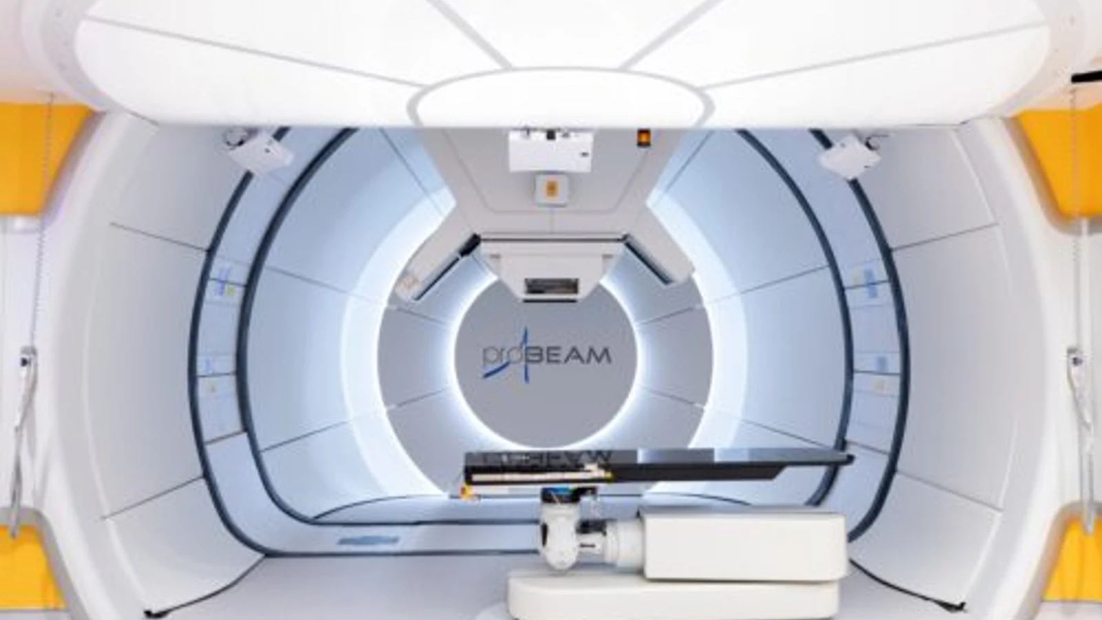
Stereotactic radio surgery for brain tumor lesions is an outpatient, non-invasive technique that directs focused radiation beams rather than a scalpel. Despite the name "radiosurgery," it involves no surgical incisions.
- Mechanism of Action: Advanced delivery platforms, such as the Leksell Gamma Knife, CyberKnife, or specialized linear accelerators (LINAC), direct hundreds of low-dose radiation beams through different entry angles toward a defined target.
- Precision Dosage: Individual beams pass through healthy brain tissue with minimal impact. However, all beams converge at a single point to deliver a concentrated, lethal radiation dose to the tumor cells, damaging their DNA and halting replication.
- Clinical Applications: SRS provides effective primary control for single or multiple brain metastases, deep vestibular schwannomas (acoustic neuromas), recurrent pituitary adenomas, and skull-base meningiomas measuring under 3 to 3.5 centimeters.
Fractionated Stereotactic Radiotherapy (SRT)
When tumors lie directly next to radiation-sensitive structures like the optic chiasm, optic nerves, or brainstem, delivering a single high dose of radiation carries a risk of permanent nerve damage. Under these conditions, radiation oncologists fractionate the total dose into multiple smaller sessions (usually 3 to 30 visits). This fractionation allows intervening healthy tissue time to repair sub-lethal DNA damage between visits while continuing to destroy tumor cells.
Systemic Chemotherapy and Targeted Molecular Therapy
Blood-brain barrier permeability remains a key challenge for medical neuro-oncologists. While many systemic chemotherapies struggle to cross from blood vessels into brain parenchyma, select agents and novel formulations achieve effective therapeutic concentrations:
- Alkylating Agents: Drugs like temozolomide, lomustine (CCNU), and carmustine (BCNU) penetrate the blood-brain barrier effectively.
- Targeted Inhibitors: Tumors harboring specific genetic variants benefit from precision therapies. For example, BRAF V600E-mutated low-grade or high-grade gliomas are treated with combined dabrafenib and trametinib regimens approved by the US FDA.
- Anti-Angiogenic Agents: Bevacizumab, a monoclonal antibody binding vascular endothelial growth factor (VEGF), reduces tumor vascularization and helps control treatment-resistant cerebral edema.
What Can Patients Expect During Recovery and Rehabilitation?
Postoperative hospital stays typically last three to seven days, followed by several weeks to months of outpatient physical, occupational, and speech therapy depending on tumor location and baseline neurological health. Recovery timelines reflect each patient's individual surgical trajectory, the surgical approach, and the brain region involved.
Recovery follows a progressive, coordinated path across clinical and home environments:
Immediate Neuro-Intensive Care Unit (NICU) Phase (Hours 0-48):
- The clinical team maintains continuous arterial line blood pressure monitoring, carries out hourly pupil and motor checks, and manages brain swelling with dexamethasone or hypertonic solutions.
- An early contrast-enhanced brain MRI is completed within 24 to 72 hours of surgery to measure the extent of resection and look for early surgical changes.
- Physical therapy staff help patients stand and take their first steps within 24 to 36 hours once intracranial stability is established.
Step-Down Inpatient Ward Phase (Days 3-7):
- Invasive monitoring lines, central venous catheters, and surgical subgaleal drains are gradually removed.
- Clinical teams taper steroid doses to avoid prolonged side effects while watching closely for rebound brain swelling.
- Physical, occupational, and speech-language therapists evaluate the patient's functional abilities to design an appropriate discharge plan.
Subacute Healing and Inpatient or Outpatient Rehabilitation (Weeks 2-8):
- Patients transition to home health programs or specialized inpatient neuro-rehabilitation facilities if they need continued help regaining functional independence.
- Care teams perform wound checks and remove scalp sutures or titanium staples around postoperative days 10 to 14.
- Temporary postoperative neurological fatigue is common during this period as the brain heals and reroutes neural pathways.
Long-Term Functional Integration (Month 3 and Beyond):
- Patients gradually return to professional work, driving, and social routines, guided by objective cognitive testing and seizure-free intervals required by local driving regulations.
- The neuro-oncology team conducts scheduled surveillance MRI scans, usually every two to four months for high-grade malignancies and every six to twelve months for stable benign lesions.
What Is the Cost of Brain Tumor Treatment at Home and Abroad?
Brain tumor surgery in the US regularly exceeds $50,000 to $150,000 without insurance, while comparable neurosurgical care in accredited international centers in Turkey or Germany typically ranges from $15,000 to $45,000. These wide variations stem from differing hospital administrative overhead, healthcare labor costs, regional surgical device pricing, and national medical cost structures.
When evaluating the overall cost of brain tumor treatment, international patients should examine all-inclusive bundled packages rather than focusing solely on isolated surgical fees. Comprehensive care plans typically incorporate:
- Advanced pre-operative neuro-navigation protocols and functional neuroimaging.
- Surgeon, assistant neurosurgeon, and specialized neuroanesthesia professional fees.
- Use of the operating suite, including intraoperative navigation, operating microscopes, and ultrasonic surgical aspirators.
- Dedicated neuro-intensive care unit (NICU) monitoring and acute surgical ward stays.
- Standard inpatient medications, including osmotic diuretics, anticonvulsants, and corticosteroids.
- Initial physical therapy, occupational assessments, and early speech-language evaluations.
Global Cost Comparison Overview
| Country / Region | Average Surgical Resection Cost (USD) | Stereotactic Radiosurgery (Gamma Knife / CyberKnife) | Typical Inpatient Stay Duration | Primary Hospital Accreditation |
|---|---|---|---|---|
| United States | $80,000-$150,000+ | $25,000-$55,000 | 3-6 days | Joint Commission / CMS |
| United Kingdom (Private) | £35,000-£65,000 ($45,000-$85,000) | £15,000-£28,000 ($19,000-$36,000) | 4-7 days | Care Quality Commission (CQC) |
| Germany | €25,000-€48,000 ($27,000-$52,000) | €10,000-€18,000 ($11,000-$19,500) | 5-8 days | KTQ / DIN EN ISO / JCI |
| Turkey | $16,000-$32,000 | $6,000-$12,000 | 4-7 days | Joint Commission International (JCI) / Licensed by Turkish MoH |
International medical travelers should evaluate prospective neurosurgical facilities by confirming:
- Institutional Accreditation: Facilities with JCI accreditation meet rigorous patient safety standards, follow strict infection prevention protocols, and use well-established surgical verification checklists.
- Neurosurgical Case Volume: High surgical volumes correlate with better clinical outcomes. Surgeons who regularly perform complex cranial resections each year generally maintain lower complication rates.
- Multidisciplinary Tumor Boards: Safe neuro-oncology care depends on regular collaboration. Look for centers where neurosurgeons, radiation oncologists, medical neuro-oncologists, neuropathologists, and neuroradiologists review complex cases together.
- Advanced Technology Suites: Leading operating theaters feature intraoperative MRI or CT, high-magnification surgical microscopes, neuro-endoscopes, and intraoperative neurophysiological monitoring (IONM) setups.
Risks and Potential Complications of Brain Tumor Surgery
Brain tumor surgery carries distinct neurological, surgical, and anesthetic risks because cranial procedures involve delicate vascular networks and complex parenchymal architecture. Neurosurgeons balance these considerations by weighing the natural progression of an untreated tumor against the anticipated complications of surgical resection.
Reported complication rates vary depending on tumor location, deep vascular involvement, and a patient's overall health:
- Focal Neurological Deficits: Postoperative deficits, such as limb weakness (hemiparesis), sensory changes, coordination loss (ataxia), or speech difficulties (aphasia), occur in roughly 5% to 15% of cranial procedures. These deficits may be temporary, improving over weeks as local swelling subsides, or they can become permanent if essential blood vessels or brain tracts are damaged.
- Intracranial Hemorrhage and Swelling: Postoperative bleeding within the resection cavity happens in approximately 1% to 3% of cases. Bleeding can abruptly raise intracranial pressure, sometimes requiring emergency re-exploration to evacuate hematomas and relieve pressure.
- Cerebrospinal Fluid (CSF) Leaks: Leakage of cerebrospinal fluid through the dura mater and wound or nasal cavity occurs in 2% to 8% of skull-base and endoscopic procedures. Left untreated, a CSF leak increases the risk of meningitis, meaning it often requires lumbar drainage or surgical repair.
- Surgical Site Infections and Meningitis: Deep intracranial infections occur in 1% to 3% of elective cranial surgeries. Care teams lower this risk by administering prophylactic IV antibiotics before incision, using strict sterile protocols, and thoroughly irrigating the surgical site.
- Seizures: Even in patients who have never had a seizure before, surgical manipulation of the cerebral cortex can trigger postoperative seizures in 4% to 10% of cases. Because of this, care teams routinely administer prophylactic anticonvulsants (such as levetiracetam) throughout the perioperative window.
- Deep Vein Thrombosis (DVT) and Pulmonary Embolism (PE): Patients with brain tumors face a higher risk of systemic blood clots due to reduced physical mobility, pro-inflammatory factors from the tumor, and long surgical procedures. Teams counter this using pneumatic compression devices and carefully timed anticoagulant therapies.
Patients must seek immediate emergency medical care if they experience high fevers, a stiff neck, clear fluid draining from the incision or nose, sudden and worsening limb weakness, speech difficulties, or new, severe seizures at any point during recovery.
Frequently Asked Questions
How much does a brain tumor surgery cost in the USA?
Brain tumor surgery in the United States typically costs between $50,000 and $150,000 for hospital and surgeon fees, with complex cases or extended neuro-ICU admissions sometimes exceeding $200,000. Uninsured international patients usually receive customized self-pay quotes that require upfront deposits covering the surgical suite, specialized neuro-navigation technology, and dedicated intensive care monitoring.
Is awake brain tumor surgery painful?
Awake brain tumor surgery is typically well tolerated and rarely painful because brain tissue lacks sensory pain receptors (nociceptors). Neurosurgeons apply targeted local and regional nerve blocks to numb the scalp, bone, and sensitive meninges, while intravenous sedation keeps the patient relaxed during the craniotomy before they are gently awakened for the mapping portion of the procedure.
Can a brain tumor return after complete surgical removal?
A brain tumor can return after complete surgical resection depending on its underlying histology, WHO grade, and microscopic infiltration. While benign Grade 1 meningiomas that are completely resected show very low recurrence rates over ten years, high-grade malignancies like glioblastomas almost always recur locally, which is why patients need continuing surveillance MRI scans alongside radiation and chemotherapy.
How long must a patient wait before flying home after brain tumor surgery?
Patients should generally avoid commercial air travel for two to four weeks following open cranial surgery. This waiting window allows air trapped in the skull (pneumocephalus) to resorb naturally; flying too soon can cause this trapped air to expand under changing airplane cabin pressures, raising intracranial pressure and causing severe headaches or other neurological complications.
How do doctors decide between stereotactic radiosurgery and open craniotomy?
Multidisciplinary neuro-oncology teams determine the best treatment strategy by evaluating tumor volume, tissue histology, physical location, and the severity of mass effect. Open craniotomy remains the preferred approach for large masses causing severe intracranial pressure and brain compression, while stereotactic radiosurgery is favored for smaller, deeply situated, surgically hard-to-reach lesions or multiple brain metastases measuring under three centimeters.
Not sure which hospital is right for your case?
Upload your medical reports and our AI will match them against the verified capabilities of every hospital in our network.
- Upload your reports (no account needed)
- Our AI matches your case with hospitals that can actually treat it
- You choose who receives your case. We present it in the hospital’s own language, and the hospital replies directly to you
Free and anonymous. Your documents stay private until you decide to send them.