

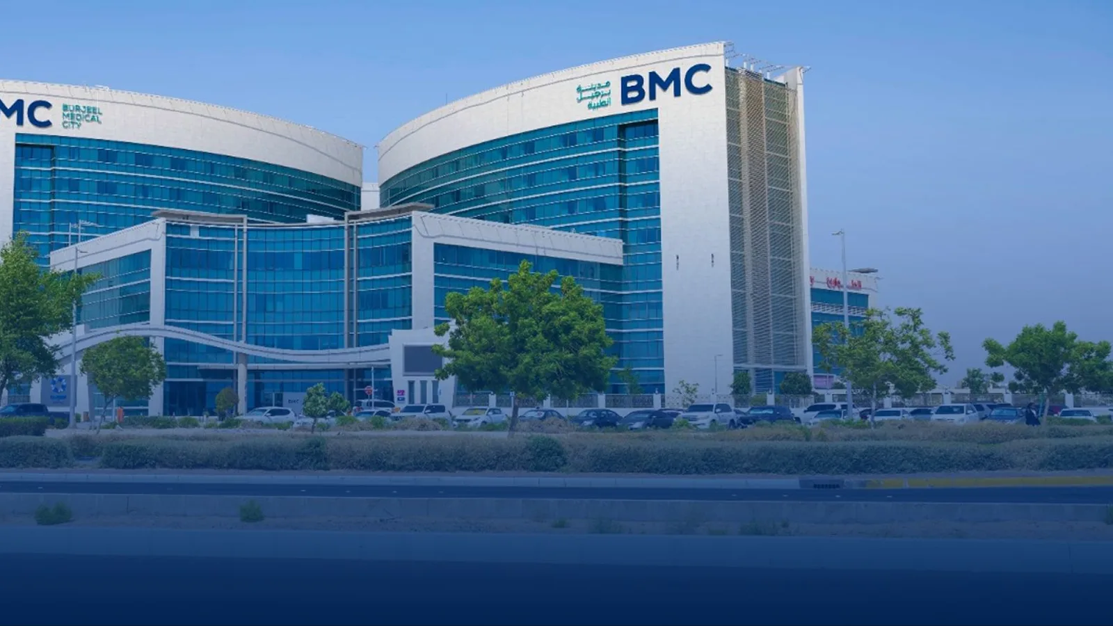

Burjeel Medical City
Abu Dhabi

- Specialties
- 29
- Departments
- 60
Not sure which hospital fits your case?
Upload your medical records and let AI match you to the right hospital.
Upload records and get matchedAbout
Burjeel Medical City is the quaternary care flagship of Burjeel Holdings, in Mohammed Bin Zayed City, Abu Dhabi. The hospital spans more than 1.2 million square feet, operates over 350 beds across more than 60 medical specialties, and works with over 200 board certified physicians. Its clinical programmes are organised into thirteen institutes and centres, among them the Burjeel Cancer Institute, the Burjeel Transplant Institute for liver and kidney transplantation, the Burjeel Orthopedic Institute with the Paley Middle East Clinic and the Al Muderis Osseointegration Clinic, the Burjeel Eye Institute, the Neuroscience Institute, the Ear Nose Throat Head and Neck Institute, the Advanced Gynecology Institute, the Maternal Fetal Institute and the Thalassemia and Sickle Cell Center. Treatment and diagnostic technology includes PET-CT and SPECT-CT, intraoperative MRI, an MR-Linac and Elekta Versa HD linear accelerators, the da Vinci Xi surgical system, a bi-plane cath lab, 3D tomosynthesis mammography and an on-site laboratory for HLA and molecular diagnostics. Emergency and critical care run around the clock with an onsite helipad, a Level III neonatal intensive care unit and a Level IV paediatric intensive care unit. The hospital holds JCI accreditation, CAP accreditation for its laboratory, ESMO designation for integrated oncology and palliative care, and CARF accreditation for rehabilitation.
Specialties
Physicians
1-12 of 143
Departments
- Acute and Trauma Care
- Advanced Gynecology Institute
- Advanced Hernia Center
- Advanced Proctology
- Advanced Rehabilitation
- Al Muderis Osseointegration Clinic
- Breast Care Center
- Burjeel Cancer Institute
- Burjeel Ear Nose Throat Head and Neck Institute
- Burjeel Eye Institute
- Burjeel Orthopedic Institute
- Burjeel Transplant Institute
- Cardiac Sciences
- Dentistry
- Dermatology and Cosmetology
- Endocrinology
- Gastroenterology
- Gastrointestinal and Bariatric Surgery
- Gastrointestinal Oncology
- General and Laparoscopic Surgery
- Gynecologic Oncology
- Hepatology
- Himmah Rehabilitation
- IFEM Endo Middle East Clinic
- Kidney Transplant
- Liver Transplant
- Maternal Fetal Institute
- Medical Oncology
- Neonatology
- Nephrology
- Neurology
- Neuroscience Institute
- Neurosurgery
- Obstetrics and General Gynecology
- Oral and Maxillofacial Surgery
- Orthopedic Oncology
- Pain Medicine
- Paley Middle East Clinic
- Pediatric Cardiology
- Pediatric Endocrinology
- Pediatric Gastroenterology
- Pediatric Nephrology
- Pediatric Neuroscience
- Pediatric Oncology
- Pediatric Pulmonology
- Pediatric Surgery
- Pediatrics
- Plastic, Reconstructive and Cosmetic Surgery
- Pulmonology
- Radiation Oncology
- Rheumatology
- Sleep Clinic
- Spine and Scoliosis
- Surgical Oncology
- Thalassemia and Sickle Cell Center
- Thoracic Surgery
- Thyroid and Parathyroid Center
- Urology
- Vascular Surgery
- Weight Management Clinic
Procedures
Bariatric and Metabolic Surgery
Cardiology and Cardiovascular Surgery
Dental, Oral and Maxillofacial Surgery
Ear, Nose and Throat
Gastroenterology
General Surgery
Gynecology and Obstetrics
Neurology and Neurosurgery
Oncology
Organ Transplantation
Orthopedics and Traumatology
Physical Medicine and Rehabilitation
Plastic and Aesthetic Surgery
Spine Surgery
International patient services
- International patient office
- Interpreter and translation services
- Visa and travel assistance
- Airport transfer
- Accommodation assistance
Technologies and equipment
Hybrid Operating Room
A hybrid operating room is a surgical theatre that combines a full operating room with advanced, built-in medical imaging in the same space. Instead of relying on portable equipment or moving a patient to a separate scanning room, the team has a fixed, high-resolution imaging system, such as a robotic angiography arm, a CT scanner or an MRI, positioned right at the operating table. This lets surgeons see detailed live pictures of the body during the procedure and combine open surgery with minimally invasive, catheter-based techniques in a single session. For the patient, it can mean a less invasive operation, immediate confirmation that the surgery worked, fewer transfers between rooms and, often, a safer option when the case is complex or high-risk.
Gait Training Robot (Adult)
A gait training robot is an advanced rehabilitation device that helps adults relearn to walk after the ability has been lost or impaired. It works on the principle of neuroplasticity: by guiding the legs through the correct walking pattern many times over, it helps the brain and muscles rebuild their connections and form new pathways, so walking becomes possible again. The patient is held safely in a support harness that takes much of the body weight, while the robot moves the legs and sensors track every step. Because it allows far more repetitions than a therapist could deliver by hand, it can speed up recovery and make early walking practice safe.
Hydrotherapy Pool
A hydrotherapy pool is a specially designed therapy pool that uses the natural buoyancy, warmth and resistance of water to make rehabilitation exercises safer and more comfortable. In the water the body becomes much lighter, so joints carry far less load and painful or weak movements become easier to perform. Guided by a physiotherapist, patients can start moving earlier and more confidently than they could on land, which makes the pool a valuable part of many rehabilitation programmes.
Excimer Laser
The excimer laser is a precise, computer-controlled laser system used to correct refractive errors of the eye, most commonly short-sightedness (myopia), long-sightedness (hyperopia) and astigmatism. It works on the cornea, the clear front surface of the eye, gently removing microscopic layers of tissue to reshape it so that light focuses correctly on the retina. It is the workhorse behind most laser vision correction procedures, where its goal is to reduce or remove a person's dependence on glasses and contact lenses. The treatment is fast, carried out under anaesthetic drops, and recovery is generally quick.
Gamma Knife
Gamma Knife is a form of stereotactic radiosurgery, a highly precise way of treating targets deep inside the head without any cut, incision or general anaesthesia. Despite its name it is not a knife and involves no surgery in the usual sense. Instead, hundreds of finely focused beams of gamma radiation are aimed from many angles so that they all meet at a single point. Each beam alone is too weak to harm the tissue it passes through, but where they converge a strong, sharply shaped dose is delivered to the target while the surrounding healthy brain is largely spared. It offers a non-invasive option for tumours and other lesions that may be difficult or risky to reach with open surgery.
Brachytherapy
Brachytherapy is a form of internal radiotherapy in which a radiation source is placed inside the body, right at or next to the tumour, rather than aimed from outside. Because the source sits so close to the target, it can deliver a high, very localised dose while the radiation falls off sharply over a short distance, sparing the healthy tissue around it. Modern systems automate this safely: thin tubes are guided to the tumour, the source travels through them along a precise plan, and it is withdrawn at the end, leaving no radiation behind in the body.
DSA Digital Subtraction Angiography
Digital subtraction angiography (DSA) is an advanced imaging method that shows the blood vessels throughout the body in fine detail. A thin catheter delivers a contrast agent into the arteries, and specialised computer processing strips away the surrounding bone and tissue so that only the vessels stand out sharply. It is used to detect vascular problems such as narrowing, aneurysm, malformation and abnormal connections in the brain, abdomen, skin and limbs. DSA is also the basis for many minimally invasive treatments, allowing a specialist to find and, in the same session, treat a vascular problem through a tiny entry point rather than open surgery.
EBUS Endobronchial Ultrasound
Endobronchial ultrasound (EBUS) is a minimally invasive method that combines bronchoscopy with ultrasound to examine the airways and the tissues and lymph nodes that surround them. A thin, flexible bronchoscope carrying a camera and a small ultrasound probe is passed through the mouth into the airways, where the probe creates real-time images of structures just beyond the airway wall, such as lymph nodes and masses that cannot be seen with a camera alone. Under this live ultrasound guidance, a fine needle can take samples for the laboratory in the same session, all without any surgical incision.
SPECT-CT
SPECT-CT is a nuclear medicine imaging method that merges single photon emission computed tomography with computed tomography in one device, capturing both how an organ functions and its anatomical structure in a single session. A low-dose radiopharmaceutical is injected and gathers in the target tissue, where a rotating gamma camera builds three-dimensional functional images while the CT scan defines the exact location within the body. By showing not just the shape of a structure but how active it is, SPECT-CT helps doctors find disease, pinpoint exactly where it sits, and plan treatment with greater confidence.
PET-CT
PET-CT is an advanced hybrid imaging method that combines positron emission tomography with computed tomography in a single scan, mapping both the metabolic activity and the anatomical structure of the body at once. A small dose of a radioactive tracer, often a glucose analogue, is injected and gathers in cells that are working harder than normal, which is typical of many tumours. Because it can show where a disease is active before it changes the shape of an organ, PET-CT is one of the most valuable tools for detecting cancer, working out how far it has spread, and checking whether treatment is working.
FibroScan
FibroScan is a non-invasive, ultrasound-based device that measures how stiff the liver is, which reflects the degree of scarring, or fibrosis, and at the same time estimates the amount of fat in the liver. It offers a fast, painless alternative to a liver biopsy, with no incision or needle, and it assesses a larger area of the organ than a tiny tissue sample would. A probe is simply placed on the skin over the liver while the patient lies down, and a numerical result is available within minutes. It is widely used to detect and follow liver conditions and to guide and monitor treatment.
Intraoperative Radiotherapy (IORT)
Intraoperative radiotherapy, or IORT, is a way of giving a single, focused dose of radiation directly to the area at highest risk during the operation itself, right after the tumour has been removed and while the patient is still asleep under anaesthesia. With the surgical wound open, the team can place the radiation applicator exactly on the tissue that needs it and gently move skin and nearby organs out of the path of the beam. The result is treatment that is aimed precisely where cancer cells are most likely to remain, while healthy tissue is shielded. For suitable patients, especially in early breast cancer, one intraoperative dose can replace or greatly shorten several weeks of radiotherapy after surgery.
ECMO
ECMO, short for extracorporeal membrane oxygenation, is an advanced life-support technology that does the work of the heart, the lungs, or both, when they are too sick to keep the body supplied with oxygen. Blood is drawn out of the body through a tube, passed through a machine that adds oxygen and removes carbon dioxide just as healthy lungs would, and then returned warmed to the body. By taking over this vital task, ECMO gives badly damaged organs a chance to rest and recover, or it keeps a patient alive while the medical team treats the underlying cause or arranges further treatment. It is used only in intensive care for the most serious, but potentially reversible, heart and lung emergencies, and it is run continuously by a specialised team around the clock. Because it demands rare expertise and resources, ECMO is available only in a small number of highly equipped centres.
EOS 3D Skeletal Imaging
EOS is a low-dose X-ray imaging system that captures the whole spine and the lower limbs in a single pass, while the patient stands naturally and bears their own weight. Two images, one from the front and one from the side, are taken at the same time, and from them the system builds a three-dimensional model of the skeleton. Because it images the body standing, it shows the skeleton in its real, load-bearing posture, which is especially important for assessing the spine and the legs. A key advantage is its very low radiation dose, much lower than standard digital X-ray, with a dedicated mode that reduces exposure even further in children.
ESWL (Shock Wave Lithotripsy)
ESWL (Extracorporeal Shock Wave Lithotripsy) is a non-surgical way to break up stones in the urinary system, such as kidney and ureter stones, without any incision. High-energy shock waves are generated outside the body and focused precisely onto the stone, shattering it into small fragments that can then pass naturally with the urine. Because nothing is inserted into the body and no cut is made, ESWL is one of the gentlest stone treatments available. It is typically a same-day procedure, after which most people return home and pass the broken fragments over the following days.
Next-Generation Sequencing (NGS)
Next-generation sequencing, often shortened to NGS, is a powerful laboratory technology that reads the genetic code of many genes at the same time, quickly and accurately. In cancer care it is the engine of precision medicine: by examining the DNA and RNA of a tumour, it reveals the specific changes driving that individual cancer and points to the treatments most likely to work against it. The same technology is used to test for inherited cancer risk and to support molecular tumour boards, where specialists match a patient's genetic profile to targeted therapies and clinical options. Because interpreting these results well requires specialised laboratories and expert teams, comprehensive genomic testing is a feature of advanced centres, and many international patients seek it out to guide a clearer, more personalised treatment plan.
Body-Weight-Supported Gait System (Anti-Gravity Treadmill)
The body-weight-supported gait system, often called an anti-gravity treadmill, is a rehabilitation device that carries part of a patient's body weight so they can walk and train safely without the fear of falling. Some systems use an overhead harness, while others enclose the lower body in an air-pressure chamber that gently lifts the patient. By reducing the load that passes through the legs and joints, it lets people start walking earlier and practice far more repetitions than they could manage on their own feet. It is used inside a wider rehabilitation programme, always under the guidance of a physiotherapist.
Elekta Versa HD Signature
Elekta Versa HD is a high-definition linear accelerator, a machine that treats cancer with precisely shaped beams of radiation from outside the body. It combines image guidance with advanced beam-shaping so the radiation oncology team can target a tumour very accurately while protecting the healthy organs and tissue around it. Because the beam can be reshaped to match the exact outline of the tumour, treatment is both more precise and more comfortable, and most people continue their normal daily routine throughout the course.
Da Vinci Robotic Surgery
The da Vinci robotic surgical system lets a surgeon perform complex operations through a few small keyhole incisions instead of one large cut. Sitting at a nearby console, the surgeon controls tiny wristed instruments and a magnified high-definition three-dimensional camera, while the robotic arms translate every hand movement into precise, steady motion inside the body. The system never acts on its own: the surgeon is in full control at all times. For patients, this minimally invasive approach often means less pain, smaller scars, less blood loss and a quicker return to normal life.
O-Arm
The O-arm is an intraoperative imaging system that rotates a full circle around the patient to produce real-time, high-resolution cross-sectional images while surgery is underway. In effect it brings a mobile CT-style scanner into the operating room, so the surgeon can see the exact position of bone, instruments and implants at the moment they are being placed, rather than relying only on images taken before the operation. It is used mainly in spine, brain and nerve, and orthopaedic trauma surgery, where it gives precise guidance for critical steps and supports greater accuracy and safety.
3 Tesla MRI
3 Tesla MRI is a high-field magnetic resonance imaging scanner that produces exceptionally detailed pictures of the inside of the body. The "3 Tesla" refers to the strength of its magnet, which is about twice that of a standard MRI scanner, and this extra power allows sharper, higher-resolution images, often in less time. Like all MRI, it uses a strong magnetic field and radio waves rather than X-rays, so there is no ionising radiation involved. It is especially valuable for examining the brain, the nervous system, joints and soft tissues, helping doctors detect and characterise problems that may be hard to see on other scans.
Whole Body MRI
Whole body MRI examines the entire body in a single session, from the head down to the upper legs and sometimes the feet, producing one connected set of detailed images. It uses a strong magnetic field and radio waves rather than X-rays, so the examination involves no ionising radiation. By covering many organs and regions at once, it offers a broad overview that can pick up disease at an early stage. This makes it useful both as a screening tool for people who want a thorough check and as a way to look at conditions that may affect more than one part of the body.
Cardiac MRI
Cardiac MRI is a non-invasive imaging method that produces remarkably detailed pictures of the heart and the large blood vessels around it. It uses a strong magnetic field and radio waves rather than X-rays, so there is no ionising radiation involved. Unlike many other heart tests, it shows not only the shape and motion of the heart but also the condition of the heart muscle itself, down to the tissue level. This makes it a powerful tool for diagnosing and guiding the treatment of a wide range of heart conditions.
Intraoperative MRI
Intraoperative MRI, also called operating room MRI, brings the power of magnetic resonance imaging directly into surgery. A specially designed scanner, integrated into the operating room, lets the surgical team obtain detailed pictures of the brain or spine while the operation is still under way. This means the surgeon can check progress during the procedure rather than waiting for a scan afterwards. Like all MRI, it uses a magnetic field and radio waves instead of X-rays, so it adds no ionising radiation. It is especially valuable for delicate tumour surgery, where seeing the result in real time can improve the outcome.
Coronary CT Angiography
Coronary CT angiography, sometimes called virtual angiography, is a computed tomography method that produces detailed images of the heart and its coronary arteries without the need for a catheter. Traditional angiography involves threading a thin tube into the arteries, but this scan does the same job from the outside, using a fast CT scanner and a contrast agent given through a vein. It clearly shows the build-up of calcium and plaque in the artery walls and any narrowing they cause. It uses X-rays, as all CT does, with techniques designed to keep the radiation dose low.
Tomosynthesis Mammography (3D Mammography)
Tomosynthesis mammography, often called 3D mammography, is an advanced form of digital mammography that builds a three-dimensional picture of the breast from a series of thin layers. Instead of a single flat image in which overlapping tissue can hide or mimic a problem, it lets the radiologist scroll through the breast slice by slice on a high-resolution screen. This makes small lesions and tumours easier to see and helps distinguish real findings from harmless overlapping tissue, which is especially valuable for screening and for women with dense breasts.
Endoscopic Ultrasound (EUS)
Endoscopic ultrasound (EUS) combines endoscopy and ultrasound in a single thin instrument, allowing the deeper layers of the digestive tract and the organs and tissues around it to be examined in detail. By placing a tiny ultrasound probe at the tip of an endoscope and guiding it inside the body, very close to the area of interest, it produces highly detailed images of structures such as the pancreas, bile ducts and nearby lymph nodes that can be hard to see from the outside. When needed, a fine needle can take a sample for the laboratory during the same procedure, all without any surgical incision.
FAPI PET/CT
FAPI PET/CT is an advanced oncological imaging technique used to detect cancer and assess how far it has spread. It uses a tracer called FAPI, short for fibroblast activation protein inhibitor, which targets the supportive cells that surround and feed many tumours rather than the sugar uptake measured by a standard PET scan. Labelled with a radioactive isotope and combined with PET and CT, it produces detailed three-dimensional images that can highlight tumours and their spread, sometimes more clearly than conventional methods, and is especially useful for cancer types that are hard to see on a routine scan.
Robotic Arm-Assisted Orthopedic Surgery
Robotic arm-assisted orthopedic surgery is a technology used mainly in knee and hip replacement to plan and carry out the operation with very high accuracy. A detailed three-dimensional plan is built from the patient's own CT scan, and during surgery a robotic arm guides the surgeon's instruments so that bone is prepared and the implant is positioned to that exact plan. The surgeon always holds and directs the instrument; the robotic arm adds steadiness and built-in limits that protect the surrounding tissue. The aim is a joint that fits and balances well, which can mean less pain and a smoother recovery.
Thulium Laser (ThuLEP)
Thulium laser is a modern surgical laser used mainly to treat an enlarged prostate, a common cause of urinary difficulty in older men. Its best-known use is the ThuLEP technique (Thulium Laser Enucleation of the Prostate), in which the overgrown inner prostate tissue is removed through the urethra with no external cut. A defining feature of the thulium laser is that it penetrates tissue only very shallowly, which allows extremely precise cutting and excellent sealing of small blood vessels as it works. The result is a minimally invasive treatment with little bleeding, a short recovery and durable relief of urinary symptoms.
NanoKnife (Irreversible Electroporation)
NanoKnife is an ablation technology, known medically as irreversible electroporation (IRE), that destroys tumour cells using short pulses of high-voltage electrical current rather than heat or cold. The current opens tiny, permanent holes in the membrane of the tumour cells, causing them to die, while the surrounding framework of tissue is largely preserved. Because it does not burn or freeze, it can be used to treat tumours that lie very close to blood vessels, bile ducts and nerves, where heat-based or cold-based methods would risk serious damage. This makes it a valuable option for selected tumours that cannot be removed by surgery.
Neuronavigation
Neuronavigation is an image-guided surgical system that acts like a precise map and GPS for the brain and spine. Using the patient's own scans built into a three-dimensional model, it shows the surgeon exactly where the instruments are inside the body in real time, so the safest, shortest route to a lesion can be planned and followed. This is especially important in the brain and spine, where targets are often small, deep and surrounded by critical nerves and blood vessels. By guiding the surgeon away from healthy structures, neuronavigation supports accuracy, smaller approaches and added safety.
Intraoperative Neuromonitoring
Intraoperative neuromonitoring is a technology that continuously checks the function of nerves and the spinal cord while brain and spine surgery is being performed. Small sensors record the electrical activity that travels along the nerves, so the surgical team gets a live warning the moment a sensitive nerve is at risk, before any lasting damage occurs. This early warning lets the surgeon adjust technique in real time and is used to help protect movement, sensation, hearing and other vital functions. It is a key safety tool in operations close to the brain, spinal cord and critical nerves.
SMILE Pro Laser
SMILE Pro is an advanced, faster version of the SMILE (Small Incision Lenticule Extraction) technique, a modern, minimally invasive laser method for correcting short-sightedness (myopia) and astigmatism. Unlike older flap-based procedures, it works through a tiny opening only a few millimetres wide, with no large corneal flap. A femtosecond laser shapes a thin disc of tissue (a lenticule) inside the cornea, which the surgeon then removes through this small incision, gently reshaping the cornea so that vision is corrected. The goal is to reduce or remove dependence on glasses and contact lenses, with a quick and comfortable recovery.
MR-Linac (MRI-Guided Radiotherapy)
MR-Linac combines a radiotherapy machine with an MRI scanner in a single device, so the radiation oncology team can watch the tumour and the soft tissue around it in real time while treatment is being delivered. Standard radiotherapy relies on planning scans taken on earlier days, but tumours and organs shift slightly from session to session and even with breathing. By seeing the target live, MR-Linac lets the team adapt the plan each day and pause or steer the beam as the tumour moves, delivering a precise dose where it is needed while better protecting healthy tissue.
Dual-Energy CT
Dual-energy CT is an advanced form of computed tomography that scans the body at two different X-ray energy levels at the same time. A standard CT uses a single energy and shows mainly the shape and density of tissues, but by comparing how structures behave at two energies, dual-energy CT can tell different materials apart far more precisely. This added information helps doctors characterise what they see, such as distinguishing one type of tissue or deposit from another, and it can often be achieved with less contrast agent and a lower radiation dose. It uses X-rays, as all CT does, but with techniques designed to keep exposure low.
Location
Burjeel Medical City, Mohammed Bin Zayed City, Abu Dhabi, United Arab Emirates
View on Google MapsAccreditations
- JCI
Not sure which hospital fits your case?
Upload your medical records and let AI match you to the right hospital.
Upload records and get matched