Proton Radiation vs Radiation Therapy: Choosing Between Proton Beam, MR-Linac, and Adaptive RT
Compare proton radiation vs radiation therapy. Learn how Bragg peak delivery, side effects, MR-Linac options, and costs differ for various cancer types.


Proton Radiation vs Radiation Therapy: Choosing Between Proton Beam, MR-Linac, and Adaptive RT
Quick Answer: Proton therapy stops precisely at tumor depth to eliminate exit radiation, making it essential for pediatric cases and tumors near critical neural structures. However, for common adult solid tumors, modern photon platforms like MR-Linac and adaptive radiation deliver comparable tumor control and side-effect profiles at a fraction of the cost.
Key Takeaways:
- Proton beams deposit maximum energy at the target (the Bragg peak) with near-zero exit dose to downstream tissue.
- Established proton superiority is concentrated in pediatric cancers, base-of-skull tumors, spinal lesions, and ocular melanoma.
- Advanced photon systems, including MR-Linac and daily adaptive radiotherapy, offer real-time soft-tissue visualization and margin reduction.
- Clinical outcomes for prostate and early-stage lung cancers show similar local control and late toxicity rates across proton and modern photon techniques.
- Total costs for proton therapy are substantially higher, requiring patients to evaluate genuine clinical benefit versus financial and logistical demands.
Radiation oncology uses targeted ionizing energy to destroy cancer cell DNA while preserving surrounding healthy organs. Selecting the optimal radiation modality involves evaluating how different particle beams-such as charged protons versus high-energy X-ray photons-interact with human tissue to balance local tumor eradication against long-term toxicity.
Understanding the Core Difference: Proton Radiation vs Radiation Therapy
Proton beam therapy delivers targeted radiation that stops precisely at a calculated tumor depth known as the Bragg peak, leaving virtually no exit dose behind the target. In contrast, modern photon radiotherapy penetrates through tumor tissue and continues through healthy structures, although advanced beam-shaping techniques significantly limit peripheral tissue exposure.
Standard radiation therapy uses high-energy X-ray photons generated by linear accelerators (Linacs). As photons enter the body, they deposit energy along their entire path: an entrance dose near the skin, peak energy absorption at a shallow depth, and a continuous exit dose as the beam exits the patient. Modern photon delivery methods, such as Intensity-Modulated Radiation Therapy (IMRT) and Volumetric Modulated Arc Therapy (VMAT), direct multiple beams from various angles to shape radiation fields around irregular tumors, distributing the exit dose across a wider volume of normal tissue.
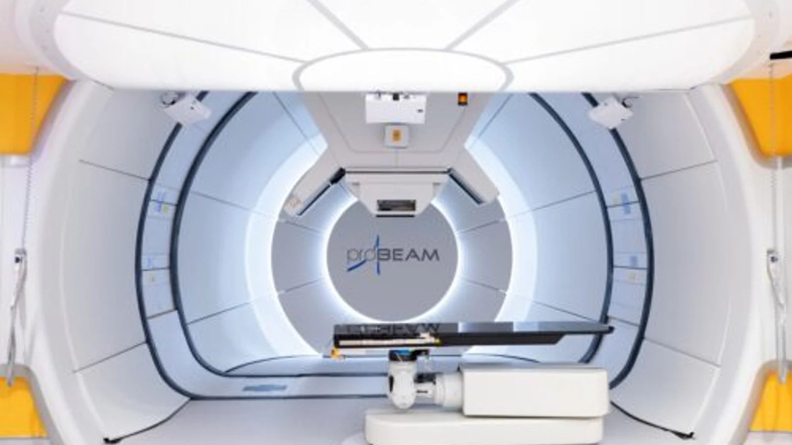
Proton radiation relies on heavy, positively charged subatomic particles accelerated by a cyclotron or synchrotron. Because protons possess mass and charge, their depth of penetration depends directly on their initial speed. As protons slow down inside tissue, their rate of energy loss rises sharply, releasing the majority of their destructive energy within a narrow window called the Bragg peak. Past this point, the radiation dose drops rapidly to zero.
When evaluating proton therapy radiotherapy alongside photon options, clinical physics teams assess pencil beam scanning (PBS) and Intensity-Modulated Proton Therapy (IMPT). PBS paints thin proton beams layer-by-layer across the tumor volume in three dimensions. This approach achieves conformality around complex shapes without exposing tissues directly behind the target.
| Feature | Conventional / Advanced Photon (IMRT / VMAT) | Magnetic Resonance Linac (MR-Linac) | Proton Beam Therapy (Pencil Beam Scanning) |
|---|---|---|---|
| Particle Type | High-energy X-ray photons | High-energy X-ray photons | Charged heavy particles (protons) |
| Exit Radiation Dose | Yes (gradual attenuation past tumor) | Yes (attenuates past tumor; tight margins) | Minimal to none (stops at the Bragg peak) |
| Real-Time Soft Tissue Imaging | On-board Cone Beam CT (low contrast) | Continuous diagnostic-grade MRI | In-room X-ray or CT (no real-time MRI) |
| Online Daily Adaptation | Available on select platforms | Standard routine before every session | Emerging, computationally complex |
| Sensitivity to Internal Motion | Low to moderate | Low (tracked directly in real time) | High (sensitive to density and gas changes) |
| Typical Treatment Course | 1 to 40 sessions (depends on cancer type) | 5 to 30 sessions | 20 to 38 sessions |
| Global Machine Availability | Over 14,000 centers globally | Growing (~150-200 centers) | Approximately 100-120 operational centers |
When Proton Therapy Is Clinically Superior: Established Indications
Proton therapy provides clear clinical advantages in pediatric malignancies, chordomas and chondrosarcomas of the skull base, ocular melanomas, and tumors directly abutting the spinal cord. In these specific conditions, eliminating exit radiation directly preserves neurological development, vision, and healthy organ function.
In pediatric oncology, developing tissues are especially vulnerable to radiation damage and secondary malignancies. Children who receive photon radiation face lifetime risks of growth disruption, neurocognitive changes, early cardiovascular disease, and secondary cancers. By eliminating the exit dose, proton therapy reduces the total integrated radiation dose to the pediatric body by up to 50-60%, making it the standard approach for medulloblastoma, ependymoma, and rhabdomyosarcoma.
For patients with a proton radiation therapy brain tumor or skull-base lesion, such as chordomas and chondrosarcomas, tumor control requires high radiation doses (often exceeding 70 Gray) close to the optic chiasm, brainstem, and temporal lobes. Proton beam therapy delivers these doses while keeping radiation to nearby critical structures within safe limits:
- Skull-Base Chordomas and Chondrosarcomas: Delivers high target doses while sparing the brainstem and cranial nerves.
- Pediatric Central Nervous System Tumors: Sparing healthy brain tissue helps prevent neurocognitive and endocrine decline.
- Ocular Melanoma: Dedicated eye proton lines achieve local tumor control while sparing the optic nerve and macula.
- Spinal and Paraspinal Sarcomas: Delivers high-dose irradiation adjacent to the spinal cord and heart.
- Re-irradiation Scenarios: When recurrent tumors develop in previously irradiated areas, the absence of an exit dose makes re-treatment feasible.
The Rise of MR-Linac and Adaptive Radiotherapy: A Viable Alternative to Protons
MR-Linac and online adaptive radiotherapy provide real-time magnetic resonance imaging and daily plan adjustments that account for internal organ movement during treatment. For localized soft-tissue tumors in the abdomen and pelvis, adaptive photon delivery often provides target precision and healthy tissue sparing comparable to proton therapy.
A key challenge with proton therapy is range uncertainty. Because protons stop abruptly based on tissue density, slight anatomical changes-such as bowel gas, bladder volume differences, or minor weight shifts-can move the Bragg peak forward or backward by several millimeters, risking marginal misses or accidental exposure of adjacent healthy tissue.
MR-Linac platforms integrate a linear accelerator with high-field (0.35T to 1.5T) magnetic resonance imaging. This setup provides high-contrast soft-tissue visualization without ionizing imaging radiation. Clinicians can track the precise boundaries of soft-tissue tumors (such as pancreatic adenocarcinoma, liver metastases, and pelvic lesions) while the patient breathes.
Online adaptive radiotherapy algorithms calculate a new, optimized photon treatment plan while the patient remains on the table, adjusting for daily organ shifts. This enables millimeter-level safety margins, significantly reducing the volume of healthy tissue exposed to photons and closing the practical gap between photon and proton distributions.
Prostate and Lung Cancers: Evaluating Proton Therapy and Radiation Therapy Side Effects
In localized prostate and lung cancers, clinical trials report similar tumor control and comparable overall toxicity rates between proton therapy and stereotactic or adaptive photon radiation. While proton therapy reduces low-dose radiation spread to distant tissues, modern photon systems like MR-Linac use tight margins that protect adjacent rectal and bladder structures.
For patients evaluating proton therapy for prostate cancer, clinical studies and randomized trials show comparable 5-year biochemical relapse-free survival between proton therapy and modern photon-based IMRT or VMAT. When assessing prostate cancer radiation therapy outcomes, both approaches demonstrate low rates of severe gastrointestinal (GI) and genitourinary (GU) toxicities when high-precision delivery and rectal spacers are used.
| Clinical Parameter | Prostate Cancer Comparison | Early-Stage Non-Small Cell Lung Cancer (NSCLC) |
|---|---|---|
| Local Tumor Control | Equivalent across Proton, MR-Linac, and VMAT | SBRT/SABR matches proton outcomes (>90%) |
| Normal Tissue Toxicity | Comparable GI/GU rates with daily image guidance | Low toxicity with SBRT; protons lower cardiac dose in Stage III |
| Motion Sensitivity | Low-to-moderate; managed with fiducials or MRI | Protons sensitive to lung density shifts; photons use 4D-CT gating |
| Standard Fractionation | 5-fraction stereotactic regimens available on both platforms | 3 to 5 fractions common for early-stage disease |
When evaluating radiation therapy side effects in non-small cell lung cancer (NSCLC), early-stage tumors treated with stereotactic body radiation therapy (SBRT/SABR) using photons achieve local control rates above 90%, matching proton results. However, in locally advanced Stage III lung cancer requiring concurrent chemoradiation, proton therapy can lower the mean radiation dose to the heart and esophagus, reducing the incidence of severe radiation pneumonitis and late cardiac complications.
Evaluating Proton Therapy Price, Insurance Coverage, and Global Availability
The overall proton therapy price typically ranges between $30,000 and $100,000 depending on treatment complexity and country, making it substantially more expensive than standard or stereotactic photon radiotherapy. Due to high installation costs, proton centers remain limited worldwide, whereas advanced photon linear accelerators are broadly accessible.
A single proton facility with multiple treatment gantries requires a capital investment exceeding $100 million due to cyclotrons, rotating gantries, and specialized concrete shielding. In contrast, advanced photon platforms (including MR-Linac units) generally cost between $3 million and $10 million.
These equipment and operational costs directly affect treatment expenses:
- United States: Proton therapy regimens typically range from $40,000 to over $120,000, whereas photon IMRT or SBRT ranges from $15,000 to $45,000.
- Europe and the UK: Private proton courses cost roughly €35,000 to €75,000 (£30,000 to £65,000), compared to €10,000 to €25,000 for adaptive photon therapy.
- Asia (e.g., Japan, South Korea): Out-of-pocket costs for international patients range from $25,000 to $50,000.
Insurance providers evaluate proton therapy against established medical necessity criteria. Approvals are standard for pediatric tumors, central nervous system chordomas, and ocular melanoma. For common adult malignancies, such as localized prostate or breast cancer, insurers typically require evidence of anatomical complexity or prior radiation before approving proton therapy over advanced photon options.
Five Questions to Ask Your Radiation Oncologist Before Choosing Your Treatment
Patients should ask their radiation oncologist: 1) What specific normal tissue sparing benefit does proton therapy offer over an adaptive photon plan for my exact tumor? 2) Does my anatomy involve motion that favors real-time MR guidance over proton sensitivity to tissue changes? 3) What are the comparative risk models for secondary malignancies based on my age? 4) How will treatment delays for proton travel affect my overall cancer timeline? 5) Does comparative trial data show improved survival or lower severe toxicity for my stage?
Use these structured questions during clinical consultations:
"Can you generate a comparative plan showing both photon (IMRT/VMAT/MR-Linac) and proton dose distributions for my anatomy?" Why to ask: Dose-volume histograms (DVHs) show whether proton therapy meaningfully reduces dose to critical organs at risk in your specific situation.
"Does my tumor move with breathing or organ filling, and how does each platform account for this?" Why to ask: Moving targets in the chest and abdomen can cause proton range errors, whereas MR-Linac provides direct real-time soft-tissue tracking.
"What is my baseline risk for secondary radiation-induced malignancies over the next 15 to 30 years?" Why to ask: Younger patients benefit more from lower whole-body low-dose radiation baths than older patients with limited lifetime secondary cancer risk.
"Will traveling to an out-of-region proton center cause treatment delays or disrupt concurrent chemotherapy regimens?" Why to ask: Unintended gaps in treatment schedules can negatively affect overall tumor control rates.
"What do phase III randomized clinical trials show for my specific cancer stage when comparing protons against advanced photons?" Why to ask: Clarifies whether proton therapy offers proven survival or toxicity benefits for your tumor type, or if modern photon radiation delivers equivalent clinical results.
Potential Risks and Treatment-Related Complications
All radiation modalities carry risks of acute and late side effects resulting from radiation exposure to healthy tissues in the treatment path.
Acute side effects develop during or shortly after treatment and occur with both proton and photon therapies:
- Fatigue: Generalized fatigue occurs across all modalities due to systemic cellular repair demands.
- Dermatologic Reactions: Skin redness, dryness, and peeling along beam entrance points can occur with both modalities, though protons deliver a slightly higher entrance dose per beam.
- Localized Mucositis and Inflammation: Depending on the treated area, temporary inflammation of the esophagus, bowel, or bladder may develop.
Late complications can appear months or years following treatment:
- Tissue Fibrosis: Radiation-induced tissue thickening can reduce local organ flexibility.
- Secondary Malignancies: Ionizing radiation carries a small lifetime risk (typically estimated under 1-3% at 10-15 years) of inducing secondary tumors. Protons reduce whole-body scatter, lowering this risk primarily in children and young adults.
- Proton Range Uncertainty Complications: If anatomical changes (such as weight loss, sinus fluid accumulation, or bowel gas) shift the Bragg peak beyond the planned margin, tissues immediately adjacent to the tumor edge can receive unintended high doses.
Radiation teams reduce these risks using daily image verification, rigid immobilization, motion management techniques (such as respiratory gating and breath-hold protocols), and verification CT scans throughout treatment.
Frequently Asked Questions
How long does a standard course of proton therapy take compared to photon treatment?
Proton therapy courses typically require daily weekday sessions over 4 to 7 weeks (20 to 35 fractions), depending on cancer type and stage. While hypofractionation (1 to 5 high-dose sessions) is standard for many photon SBRT and MR-Linac protocols, proton centers are also adopting 5-fraction regimens for select prostate and lung cancer cases.
Can a patient switch between proton beam therapy and photon radiotherapy during treatment?
Switching modalities mid-treatment is possible but technically complex, requiring complete replanning and biological dose recalculations. Radiation oncologists generally avoid switching unless equipment access is interrupted or significant anatomical changes make continuing on the initial system clinically unsuitable.
Why is proton therapy sensitive to weight changes and anatomical shifts?
Because the proton Bragg peak stops at a depth determined by tissue electron density, any change in the beam's path-such as weight loss, muscle changes, fluid buildup, or bowel gas-alters where the beam deposits its maximum dose. Photon beams pass through tissue continuously, making their energy distribution less sensitive to minor day-to-day density variations.
Is proton therapy covered by international health insurance for adult tumors?
Coverage depends on policy terms, the patient's country of origin, and the specific clinical diagnosis. While international insurers routinely approve proton therapy for pediatric, central nervous system, and skull-base tumors, common adult cancers (like localized prostate or breast cancer) typically require detailed clinical review and documentation that photon therapy would exceed normal tissue tolerance limits.
Not sure which hospital is right for your case?
Upload your medical reports and our AI will match them against the verified capabilities of every hospital in our network.
- Upload your reports (no account needed)
- Our AI matches your case with hospitals that can actually treat it
- You choose who receives your case. We present it in the hospital’s own language, and the hospital replies directly to you
Free and anonymous. Your documents stay private until you decide to send them.