


Neurology and Neurosurgery
Istanbul

Neurology and Neurosurgery care in Istanbul is available at 12 hospitals in the Voumed network, with 3 related treatments.
Neurology and neurosurgery are the medical and surgical branches that care for the brain, spinal cord, nerves and muscles. Neurology leads on diagnosis and non-surgical treatment of conditions such as stroke, epilepsy, migraine, multiple sclerosis, Parkinson's disease and dementia, while neurosurgery operates on the brain and spine when surgery is the better path. Together they cover everything from a sudden stroke to a slow-growing brain tumour or a worn disc pressing on a nerve. Patients often travel abroad for this care because complex brain and spine work depends on experienced teams, advanced imaging and modern operating technology, and because a fast, coordinated assessment can make a real difference to the outcome.
At a glance
- Sub-specialties
- stroke and cerebrovascular neurology, epilepsy, movement disorders, neuro-oncology, spinal neurosurgery, functional neurosurgery, neuromuscular disease
- Common procedures
- brain tumour surgery, spinal and disc surgery, deep brain stimulation, epilepsy surgery, aneurysm and vascular treatment, clot removal for stroke
- Common reasons to travel
- experienced surgical teams, advanced imaging and navigation technology, rapid diagnosis and a multidisciplinary plan
- Typical hospital stay
- day case or 1 to 2 nights for minor procedures, several days to about a week for major brain or spine surgery
- Anaesthesia
- general for most operations, with some functional and awake procedures using sedation and local anaesthesia by design
- Typical first step
- a neurological consultation with imaging review, so the cause is understood before any treatment is planned
Not sure which hospital fits your case?
Upload your medical records and let AI match you to the right hospital.
Upload records and get matchedAvailable at these hospitals
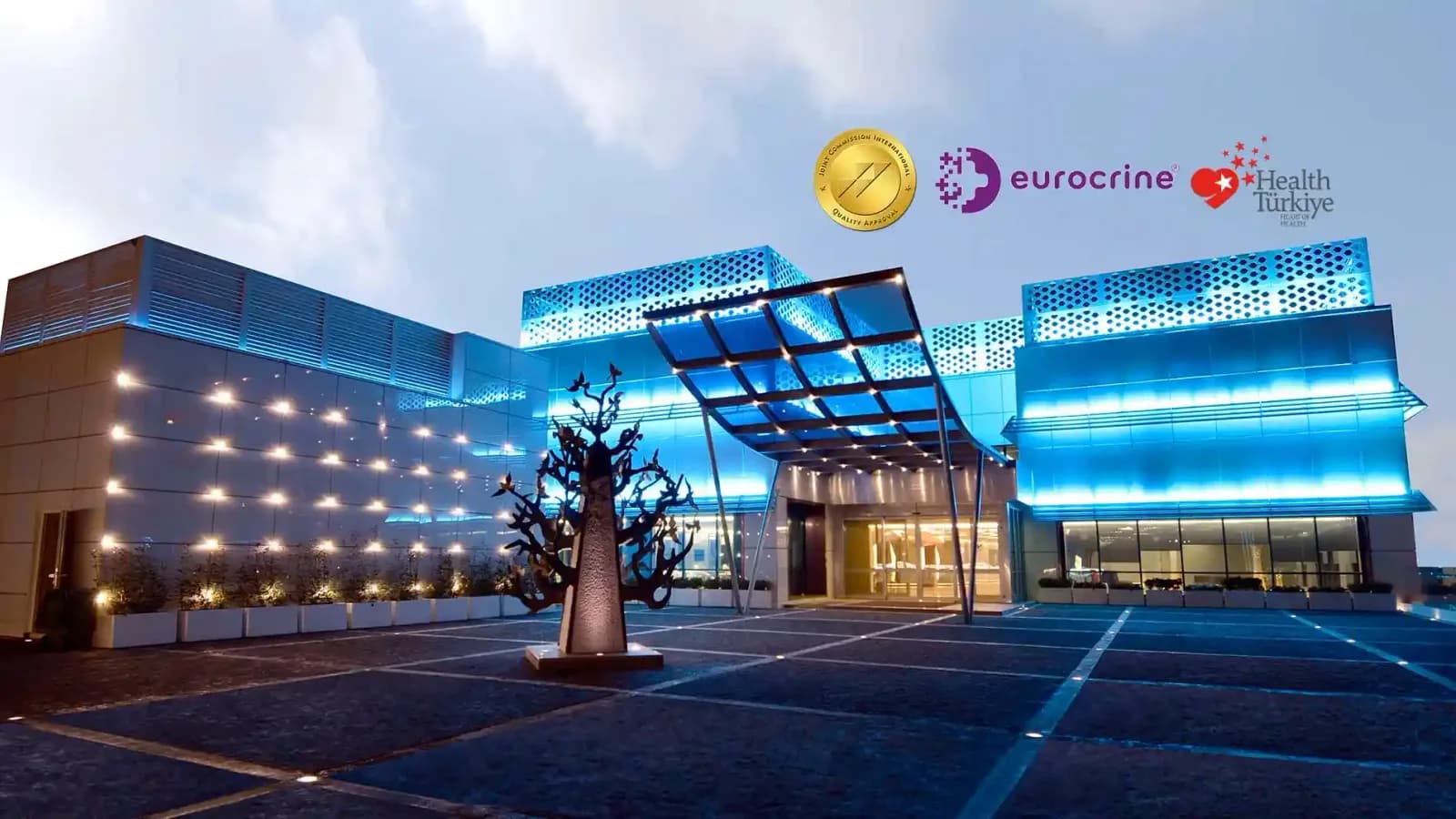

Liv Hospital Ulus
 istanbul

- Specialties
- 24

Liv Hospital Vadistanbul
istanbul
- Specialties
- 24


Acıbadem Maslak Hospital
istanbul
- Specialties
- 29

Liv Hospital Topkapı
istanbul
- Specialties
- 19

Acıbadem Altunizade Hospital
istanbul
- Specialties
- 28

Acıbadem Ataşehir Hospital
istanbul
- Specialties
- 29

Liv Hospital Bahçeşehir
istanbul
- Specialties
- 24
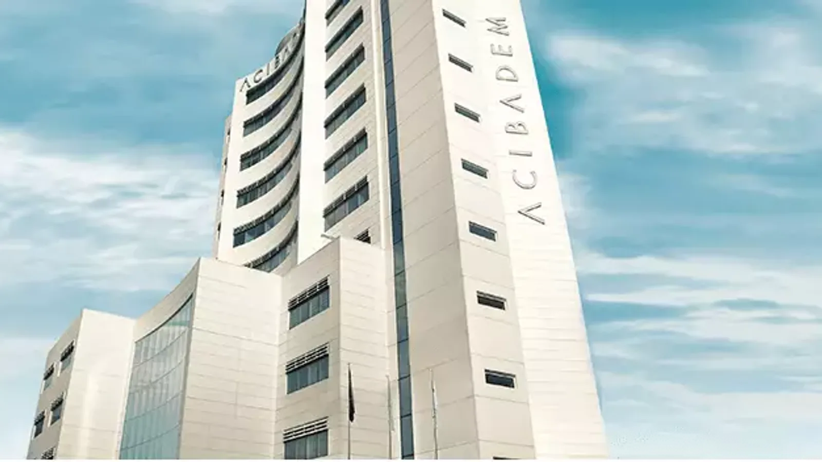
Acıbadem Fulya Hospital
istanbul
- Specialties
- 23


Memorial Bahçelievler Hospital
istanbul
- Specialties
- 31
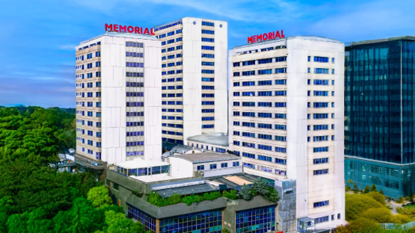
Memorial Şişli Hospital
istanbul
- Specialties
- 29
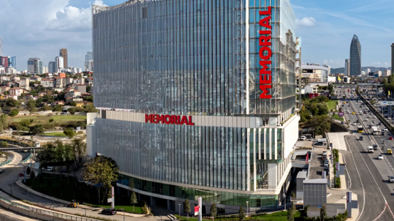
Memorial Göztepe Hospital
istanbul
- Specialties
- 25
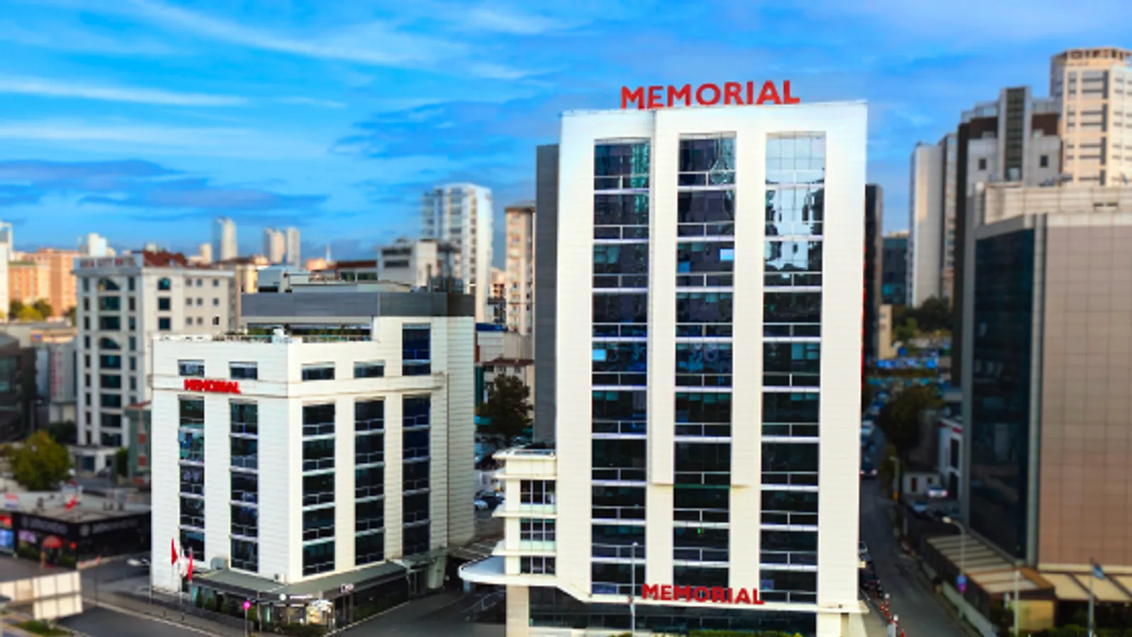
Memorial Ataşehir Hospital
istanbul
- Specialties
- 28
Procedures
Technologies and equipment
CyberKnife M6
CyberKnife M6 is a robotic system for stereotactic radiosurgery and stereotactic body radiotherapy (SBRT). Despite the name, there is no knife and no cutting. A small linear accelerator sits on a computer-guided robotic arm and delivers many thin beams of focused radiation from hundreds of angles. The beams converge on the tumour with sub-millimetre accuracy, so a high dose reaches the target while nearby healthy tissue is spared. Imaging during treatment tracks the tumour continuously, and a motion-synchronisation feature follows targets that move with breathing, such as those in the lung or liver. Treatment is non-invasive and painless, needs no rigid head frame, and is usually given as an outpatient over one to five sessions. The decision is always made individually by the radiation oncology team.
ROSA Robotic Surgical Assistant
ROSA is a robotic surgical assistant used in knee replacement and brain surgery to plan and carry out delicate operations with very high accuracy. It is used in two main areas: joint replacement, where it guides bone preparation and implant placement, and brain surgery, where it helps reach precise targets deep in the brain. In both settings the system turns the patient's own scans into a detailed three-dimensional map, and a steady robotic arm then follows the plan the surgeon has set. The surgeon remains fully in control; ROSA adds precision and stability rather than acting on its own.
Gamma Knife
Gamma Knife is a form of stereotactic radiosurgery, a highly precise way of treating targets deep inside the head without any cut, incision or general anaesthesia. Despite its name it is not a knife and involves no surgery in the usual sense. Instead, hundreds of finely focused beams of gamma radiation are aimed from many angles so that they all meet at a single point. Each beam alone is too weak to harm the tissue it passes through, but where they converge a strong, sharply shaped dose is delivered to the target while the surrounding healthy brain is largely spared. It offers a non-invasive option for tumours and other lesions that may be difficult or risky to reach with open surgery.
Hybrid Operating Room
A hybrid operating room is a surgical theatre that combines a full operating room with advanced, built-in medical imaging in the same space. Instead of relying on portable equipment or moving a patient to a separate scanning room, the team has a fixed, high-resolution imaging system, such as a robotic angiography arm, a CT scanner or an MRI, positioned right at the operating table. This lets surgeons see detailed live pictures of the body during the procedure and combine open surgery with minimally invasive, catheter-based techniques in a single session. For the patient, it can mean a less invasive operation, immediate confirmation that the surgery worked, fewer transfers between rooms and, often, a safer option when the case is complex or high-risk.
O-Arm
The O-arm is an intraoperative imaging system that rotates a full circle around the patient to produce real-time, high-resolution cross-sectional images while surgery is underway. In effect it brings a mobile CT-style scanner into the operating room, so the surgeon can see the exact position of bone, instruments and implants at the moment they are being placed, rather than relying only on images taken before the operation. It is used mainly in spine, brain and nerve, and orthopaedic trauma surgery, where it gives precise guidance for critical steps and supports greater accuracy and safety.
3 Tesla MRI
3 Tesla MRI is a high-field magnetic resonance imaging scanner that produces exceptionally detailed pictures of the inside of the body. The "3 Tesla" refers to the strength of its magnet, which is about twice that of a standard MRI scanner, and this extra power allows sharper, higher-resolution images, often in less time. Like all MRI, it uses a strong magnetic field and radio waves rather than X-rays, so there is no ionising radiation involved. It is especially valuable for examining the brain, the nervous system, joints and soft tissues, helping doctors detect and characterise problems that may be hard to see on other scans.
Intraoperative MRI
Intraoperative MRI, also called operating room MRI, brings the power of magnetic resonance imaging directly into surgery. A specially designed scanner, integrated into the operating room, lets the surgical team obtain detailed pictures of the brain or spine while the operation is still under way. This means the surgeon can check progress during the procedure rather than waiting for a scan afterwards. Like all MRI, it uses a magnetic field and radio waves instead of X-rays, so it adds no ionising radiation. It is especially valuable for delicate tumour surgery, where seeing the result in real time can improve the outcome.
Functional MRI (fMRI)
Functional MRI, often shortened to fMRI, is a special kind of magnetic resonance imaging that maps activity in the brain rather than just its structure. While a standard MRI shows the shape of the brain, fMRI reveals which regions switch on when a person performs a task such as speaking, moving a hand or sensing touch. It does this by detecting tiny changes in blood flow and oxygen that follow brain activity, all without ionising radiation. This makes it an invaluable planning tool before surgery or other treatments near important parts of the brain.
Neuronavigation
Neuronavigation is an image-guided surgical system that acts like a precise map and GPS for the brain and spine. Using the patient's own scans built into a three-dimensional model, it shows the surgeon exactly where the instruments are inside the body in real time, so the safest, shortest route to a lesion can be planned and followed. This is especially important in the brain and spine, where targets are often small, deep and surrounded by critical nerves and blood vessels. By guiding the surgeon away from healthy structures, neuronavigation supports accuracy, smaller approaches and added safety.
Intraoperative Neuromonitoring
Intraoperative neuromonitoring is a technology that continuously checks the function of nerves and the spinal cord while brain and spine surgery is being performed. Small sensors record the electrical activity that travels along the nerves, so the surgical team gets a live warning the moment a sensitive nerve is at risk, before any lasting damage occurs. This early warning lets the surgeon adjust technique in real time and is used to help protect movement, sensation, hearing and other vital functions. It is a key safety tool in operations close to the brain, spinal cord and critical nerves.
Not sure which hospital fits your case?
Upload your medical records and let AI match you to the right hospital.
Upload records and get matched