Oncology
Istanbul
Oncology care in Istanbul is available at 12 hospitals in the Voumed network, with 3 related treatments.
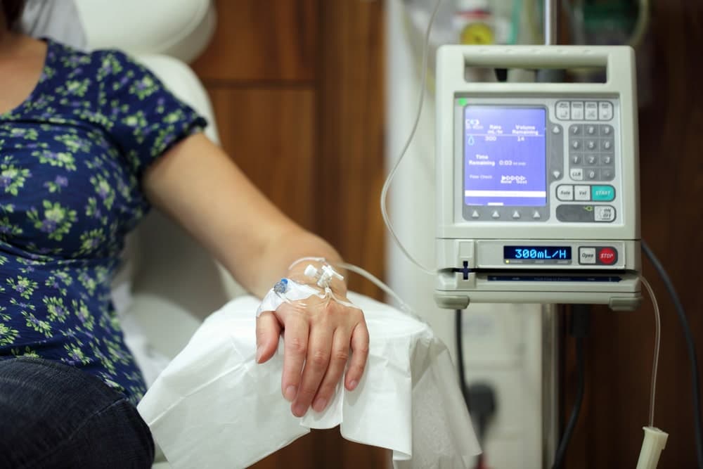
Oncology is the branch of medicine that diagnoses, treats and follows cancer, the group of diseases in which abnormal cells grow and can spread through the body. Modern cancer care is a team effort: medical oncology uses drug treatments such as chemotherapy, targeted therapy and immunotherapy, radiation oncology treats with precisely focused radiation, and surgical oncology removes tumours, while pathology, radiology, nuclear medicine and supportive care knit the plan together. Because no single specialist sees the whole picture alone, the most important decisions are usually made by a multidisciplinary tumour board that agrees one personalised plan. Patients often travel abroad for cancer care to reach experienced cancer teams, advanced radiotherapy and imaging, and timely access to treatment, all coordinated under one roof.
At a glance
- Sub-specialties
- medical oncology, radiation oncology, surgical oncology, haemato-oncology, nuclear medicine, interventional oncology
- Common treatments
- chemotherapy, targeted therapy and immunotherapy, radiotherapy, cancer surgery, ablation and artery directed therapies, bone marrow transplant
- Common reasons to travel
- experienced multidisciplinary cancer teams, advanced radiotherapy and PET imaging, timely access and second opinions
- Typical hospital stay
- many treatments are outpatient or a day case, with longer admission for major surgery or transplant
- Anaesthesia
- none for most drug and radiation treatment, local for some procedures, general for cancer surgery
- Typical first step
- review of pathology and scans, accurate staging, and a plan agreed by the tumour board
Not sure which hospital fits your case?
Upload your medical records and let AI match you to the right hospital.
Upload records and get matchedAvailable at these hospitals

Liv Hospital Vadistanbul
![]() istanbul
istanbul
- Specialties
- 24

Acıbadem Maslak Hospital
![]() istanbul
istanbul
- Specialties
- 29

Liv Hospital Topkapı
![]() istanbul
istanbul
- Specialties
- 18
Liv Hospital Ulus
![]() istanbul
istanbul
- Specialties
- 24

Acıbadem Ataşehir Hospital
![]() istanbul
istanbul
- Specialties
- 29

Acıbadem Altunizade Hospital
![]() istanbul
istanbul
- Specialties
- 28

Acıbadem Fulya Hospital
![]() istanbul
istanbul
- Specialties
- 22

Liv Hospital Bahçeşehir
![]() istanbul
istanbul
- Specialties
- 24

Memorial Bahçelievler Hospital
![]() istanbul
istanbul
- Specialties
- 31
Memorial Şişli Hospital
![]() istanbul
istanbul
- Specialties
- 29
Memorial Göztepe Hospital
![]() istanbul
istanbul
- Specialties
- 24

Memorial Ataşehir Hospital
![]() istanbul
istanbul
- Specialties
- 28
Procedures
Technologies and equipment
CyberKnife M6
CyberKnife M6 is a robotic system for stereotactic radiosurgery and stereotactic body radiotherapy (SBRT). Despite the name, there is no knife and no cutting. A small linear accelerator sits on a computer-guided robotic arm and delivers many thin beams of focused radiation from hundreds of angles. The beams converge on the tumour with sub-millimetre accuracy, so a high dose reaches the target while nearby healthy tissue is spared. Imaging during treatment tracks the tumour continuously, and a motion-synchronisation feature follows targets that move with breathing, such as those in the lung or liver. Treatment is non-invasive and painless, needs no rigid head frame, and is usually given as an outpatient over one to five sessions. The decision is always made individually by the radiation oncology team.
TomoTherapy
TomoTherapy is an image-guided radiotherapy system that delivers radiation in a continuous spiral as the treatment ring rotates around the patient, much like a CT scanner. Built-in CT imaging lets the radiation oncology team confirm the tumour's exact position before every session, and the beam is divided into many small beamlets that paint the dose precisely onto the target. This slice-by-slice approach is well suited to complex or unusually shaped tumours and to large or long treatment areas, while keeping nearby healthy organs better protected.
Next-Generation Sequencing (NGS)
Next-generation sequencing, often shortened to NGS, is a powerful laboratory technology that reads the genetic code of many genes at the same time, quickly and accurately. In cancer care it is the engine of precision medicine: by examining the DNA and RNA of a tumour, it reveals the specific changes driving that individual cancer and points to the treatments most likely to work against it. The same technology is used to test for inherited cancer risk and to support molecular tumour boards, where specialists match a patient's genetic profile to targeted therapies and clinical options. Because interpreting these results well requires specialised laboratories and expert teams, comprehensive genomic testing is a feature of advanced centres, and many international patients seek it out to guide a clearer, more personalised treatment plan.
Brachytherapy
Brachytherapy is a form of internal radiotherapy in which a radiation source is placed inside the body, right at or next to the tumour, rather than aimed from outside. Because the source sits so close to the target, it can deliver a high, very localised dose while the radiation falls off sharply over a short distance, sparing the healthy tissue around it. Modern systems automate this safely: thin tubes are guided to the tumour, the source travels through them along a precise plan, and it is withdrawn at the end, leaving no radiation behind in the body.
Varian Edge
Varian Edge is an advanced radiosurgery platform, a high-precision machine that treats tumours with very focused beams of radiation delivered from outside the body, without any cutting. It is built for stereotactic radiosurgery and stereotactic body radiotherapy, two techniques that concentrate a high dose of radiation onto the exact shape of a target while protecting the healthy tissue around it. Image guidance and real-time motion management track tiny patient or tumour movement and keep the beam on target with sub-millimetre accuracy. Treatment is painless, needs no rigid head frame for brain targets, and is usually completed in a small number of short outpatient sessions. The radiation oncology team always plans each case individually.
Ethos Adaptive Radiotherapy
Ethos is an adaptive radiotherapy system that uses artificial intelligence to tailor each cancer treatment to the patient's anatomy on the very day it is delivered. Bodies change a little from session to session: a tumour can shrink, organs shift, the bladder or bowel fill differently. Ethos takes a fresh image at the start of every session, detects these changes and, with AI support, can generate an updated plan in minutes rather than the hours such replanning would normally take, so the dose stays focused on the tumour while better protecting healthy organs.
SPECT-CT
SPECT-CT is a nuclear medicine imaging method that merges single photon emission computed tomography with computed tomography in one device, capturing both how an organ functions and its anatomical structure in a single session. A low-dose radiopharmaceutical is injected and gathers in the target tissue, where a rotating gamma camera builds three-dimensional functional images while the CT scan defines the exact location within the body. By showing not just the shape of a structure but how active it is, SPECT-CT helps doctors find disease, pinpoint exactly where it sits, and plan treatment with greater confidence.
Scalp Cooling System
A scalp cooling system is a supportive technology that helps reduce hair loss during chemotherapy, one of the side effects patients often find most distressing. The patient wears a snug cap that gently chills the scalp before, during and after the chemotherapy session. Cooling narrows the small blood vessels in the scalp and slows the activity of the hair follicles, so that less of the chemotherapy drug reaches them and they are less affected. For many people, this helps keep more of their own hair through treatment, which can make a meaningful difference to confidence and daily life.
Intraoperative Radiotherapy (IORT)
Intraoperative radiotherapy, or IORT, is a way of giving a single, focused dose of radiation directly to the area at highest risk during the operation itself, right after the tumour has been removed and while the patient is still asleep under anaesthesia. With the surgical wound open, the team can place the radiation applicator exactly on the tissue that needs it and gently move skin and nearby organs out of the path of the beam. The result is treatment that is aimed precisely where cancer cells are most likely to remain, while healthy tissue is shielded. For suitable patients, especially in early breast cancer, one intraoperative dose can replace or greatly shorten several weeks of radiotherapy after surgery.
PET-CT
PET-CT is an advanced hybrid imaging method that combines positron emission tomography with computed tomography in a single scan, mapping both the metabolic activity and the anatomical structure of the body at once. A small dose of a radioactive tracer, often a glucose analogue, is injected and gathers in cells that are working harder than normal, which is typical of many tumours. Because it can show where a disease is active before it changes the shape of an organ, PET-CT is one of the most valuable tools for detecting cancer, working out how far it has spread, and checking whether treatment is working.
TrueBeam STx
TrueBeam STx is an advanced linear accelerator, a machine that delivers external radiotherapy to treat cancer with very high precision. It shapes powerful radiation beams to match the exact size and shape of a tumour and aims them from many angles, so that a strong dose reaches the target while nearby healthy tissue and organs receive as little as possible. Because it tracks the target and can account for movement such as breathing, it is accurate to within millimetres. This makes it suitable both for conventional, daily radiotherapy and for advanced focused techniques that treat a tumour in only a few sessions. The treatment is non-invasive and painless, with nothing entering the body.
Elekta Versa HD Signature
Elekta Versa HD is a high-definition linear accelerator, a machine that treats cancer with precisely shaped beams of radiation from outside the body. It combines image guidance with advanced beam-shaping so the radiation oncology team can target a tumour very accurately while protecting the healthy organs and tissue around it. Because the beam can be reshaped to match the exact outline of the tumour, treatment is both more precise and more comfortable, and most people continue their normal daily routine throughout the course.
Whole Body MRI
Whole body MRI examines the entire body in a single session, from the head down to the upper legs and sometimes the feet, producing one connected set of detailed images. It uses a strong magnetic field and radio waves rather than X-rays, so the examination involves no ionising radiation. By covering many organs and regions at once, it offers a broad overview that can pick up disease at an early stage. This makes it useful both as a screening tool for people who want a thorough check and as a way to look at conditions that may affect more than one part of the body.
Digital Mammography
Digital mammography is a low-dose X-ray method used to screen for and detect breast cancer at an early stage. It captures very high-resolution digital images of the breast that a radiologist can examine and enhance on screen, revealing small nodules, masses and tiny specks of calcium that may not be felt or seen on other tests. Because it can find changes long before they cause symptoms, it is the cornerstone of breast cancer screening and one of the most effective tools for catching the disease when it is most treatable.
Tomosynthesis Mammography (3D Mammography)
Tomosynthesis mammography, often called 3D mammography, is an advanced form of digital mammography that builds a three-dimensional picture of the breast from a series of thin layers. Instead of a single flat image in which overlapping tissue can hide or mimic a problem, it lets the radiologist scroll through the breast slice by slice on a high-resolution screen. This makes small lesions and tumours easier to see and helps distinguish real findings from harmless overlapping tissue, which is especially valuable for screening and for women with dense breasts.
Endoscopic Ultrasound (EUS)
Endoscopic ultrasound (EUS) combines endoscopy and ultrasound in a single thin instrument, allowing the deeper layers of the digestive tract and the organs and tissues around it to be examined in detail. By placing a tiny ultrasound probe at the tip of an endoscope and guiding it inside the body, very close to the area of interest, it produces highly detailed images of structures such as the pancreas, bile ducts and nearby lymph nodes that can be hard to see from the outside. When needed, a fine needle can take a sample for the laboratory during the same procedure, all without any surgical incision.
FAPI PET/CT
FAPI PET/CT is an advanced oncological imaging technique used to detect cancer and assess how far it has spread. It uses a tracer called FAPI, short for fibroblast activation protein inhibitor, which targets the supportive cells that surround and feed many tumours rather than the sugar uptake measured by a standard PET scan. Labelled with a radioactive isotope and combined with PET and CT, it produces detailed three-dimensional images that can highlight tumours and their spread, sometimes more clearly than conventional methods, and is especially useful for cancer types that are hard to see on a routine scan.
HIFU for Prostate Cancer
HIFU (High Intensity Focused Ultrasound) is a non-surgical, targeted treatment for prostate cancer that destroys diseased tissue using precisely focused sound waves, without any cut to the body. A probe placed in the back passage delivers focused ultrasound energy that heats and destroys only the cancerous part of the prostate, while sparing the healthy tissue and nearby structures as much as possible. Because it is so targeted, HIFU aims to treat the cancer while protecting urinary control and sexual function. It is mainly used for early, localised prostate cancer and for men who want a minimally invasive option.
NanoKnife (Irreversible Electroporation)
NanoKnife is an ablation technology, known medically as irreversible electroporation (IRE), that destroys tumour cells using short pulses of high-voltage electrical current rather than heat or cold. The current opens tiny, permanent holes in the membrane of the tumour cells, causing them to die, while the surrounding framework of tissue is largely preserved. Because it does not burn or freeze, it can be used to treat tumours that lie very close to blood vessels, bile ducts and nerves, where heat-based or cold-based methods would risk serious damage. This makes it a valuable option for selected tumours that cannot be removed by surgery.
Aiforia AI Pathology
Aiforia is an artificial intelligence assisted pathology software that helps doctors analyse tissue samples taken during a biopsy or surgery. After a sample is placed on a glass slide and scanned into a high-resolution digital image, the software uses trained AI to measure and highlight features in the tissue, supporting the pathologist who makes the diagnosis. It carries a CE-IVD marking for in vitro diagnostic use and is applied in cancers such as breast, prostate, lung and skin. Importantly, it is a decision-support tool: it assists and adds consistency, but the pathologist remains responsible for the final diagnosis.
MR-Linac (MRI-Guided Radiotherapy)
MR-Linac combines a radiotherapy machine with an MRI scanner in a single device, so the radiation oncology team can watch the tumour and the soft tissue around it in real time while treatment is being delivered. Standard radiotherapy relies on planning scans taken on earlier days, but tumours and organs shift slightly from session to session and even with breathing. By seeing the target live, MR-Linac lets the team adapt the plan each day and pause or steer the beam as the tumour moves, delivering a precise dose where it is needed while better protecting healthy tissue.
512-Slice CT
512-slice CT is a very fast, high-detail computed tomography scanner that captures a large number of thin image slices with each rotation. By gathering so much information so quickly, it can build detailed three-dimensional pictures of the body in a single short breath-hold. This speed is especially valuable for imaging moving organs such as the heart, where a fast scan freezes motion and produces sharp images. It uses X-rays, like all CT, but modern scanners of this kind are designed to keep the radiation dose as low as possible.
Dual-Energy CT
Dual-energy CT is an advanced form of computed tomography that scans the body at two different X-ray energy levels at the same time. A standard CT uses a single energy and shows mainly the shape and density of tissues, but by comparing how structures behave at two energies, dual-energy CT can tell different materials apart far more precisely. This added information helps doctors characterise what they see, such as distinguishing one type of tissue or deposit from another, and it can often be achieved with less contrast agent and a lower radiation dose. It uses X-rays, as all CT does, but with techniques designed to keep exposure low.
Not sure which hospital fits your case?
Upload your medical records and let AI match you to the right hospital.
Upload records and get matched